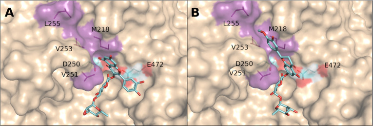
Figure 7.
Active site of αRβG II with bound rutin and hesperidin as good substrates. Bound rutin (A) or hesperidin (B) were modeled into the active site, highlighting the hydrophobic residues in the +1 subsite (magenta) that participated in the binding. The presumable catalytic nucleophile Asp250 and acid/base catalyst Glu472 are also depicted.88 The structure of αRβG II was obtained by homology modeling using MODELER153 and the PDB 4I8D. Docking was performed using Autodock4.154

