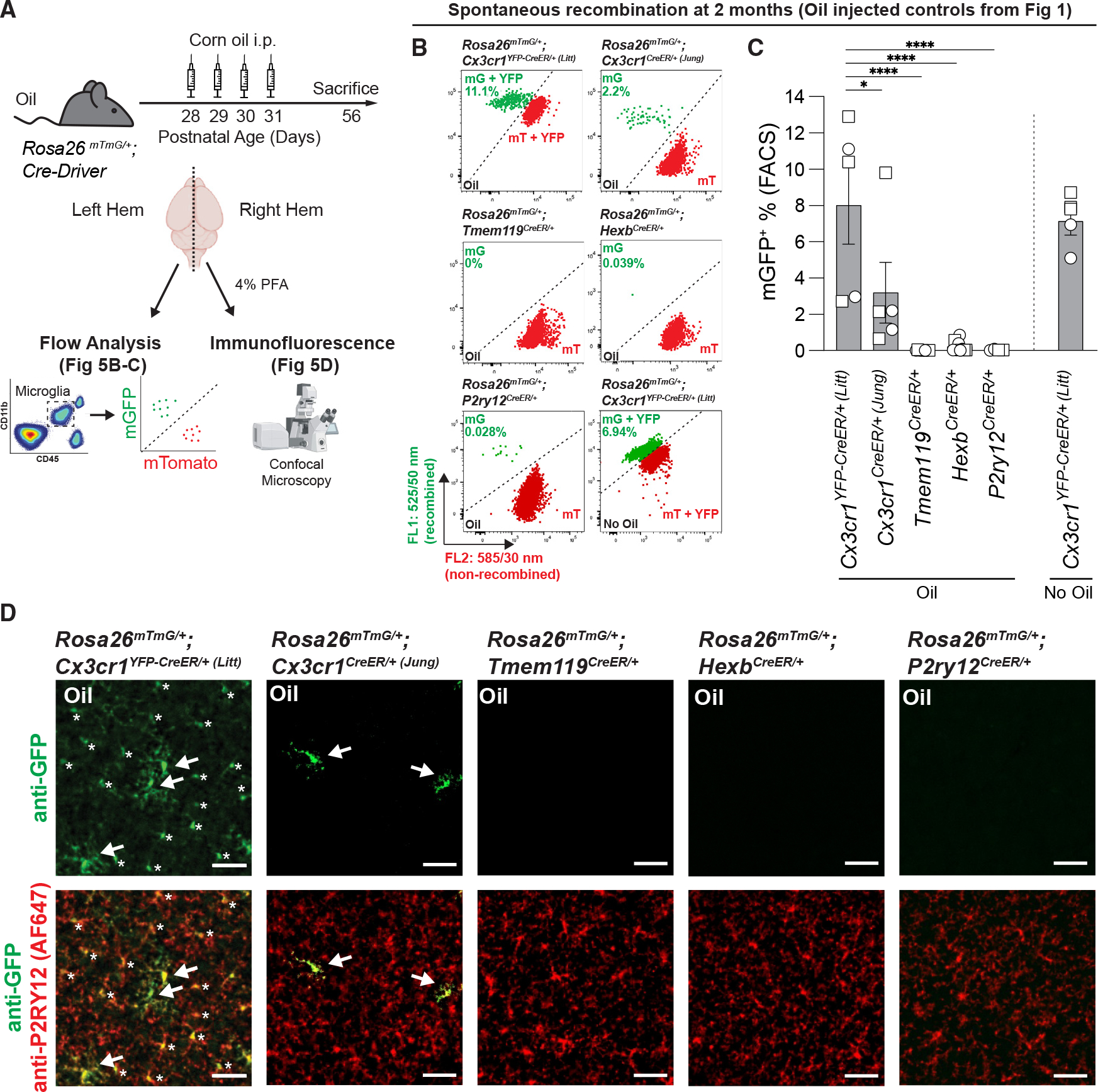
Figure 5. Spontaneous recombination of Rosa26mTmG in microglial CreER lines.

(A) Diagram of experimental protocol used to assess spontaneous Cre/loxP recombination of Rosa26mTmG/+ in microglia by flow cytometry and immunofluorescence.
(B) Representative flow cytometry results show the percentage of recombined mGFP+ (mG) and mTomato+ (mT) microglia from individual animals from each group from Figure 1 and uninjected (no oil) Cx3cr1YFP–CreER/+ (Litt) mice.
(C) Quantification of the percentage of recombined mGFP+ microglia shows increased spontaneous recombination of the Rosa26mTmG allele in the Cx3cr1YFP–CreER (Litt) line compared with the Cx3cr1CreER (Jung), Tmem119CreER, HexbCreER, and P2ry12CreER lines (one-way ANOVA with Tukey’s post hoc test; n = 5 Cx3cr1YFP–CreER/+ (Litt), 5 Cx3cr1CreER/+ (Jung), 8 Tmem119CreER/+, 9 HexbCreER/+, and 4 P2ry12CreER/+ mice; *p < 0.05, ****p < 0.0001).
(D) Representative immunofluorescent images of brain sections from right hemispheres of oil-injected mice used for flow cytometry analysis in (B) and (C). Sections were immunolabeled for anti-P2RY12 (AF647 pseudo-colored red) to identify microglia and anti-GFP (green) to identify recombined cells. The number of recombined mGFP+ microglia (white arrows) matches the results observed by flow cytometry. In the Cx3cr1YFP–CreER/+ (Litt) line, the soma of non-recombined microglia are also immunolabeled by anti-GFP because of the constitutive expression of YFP (asterisks), but it can be distinguished from recombined mGFP+ microglia by fluorescence intensity and membrane labeling. Scale bars, 50 μm.
All data are presented as mean ± SEM. Individual data points indicate males (squares) and females (circles). See also Figure S5.
