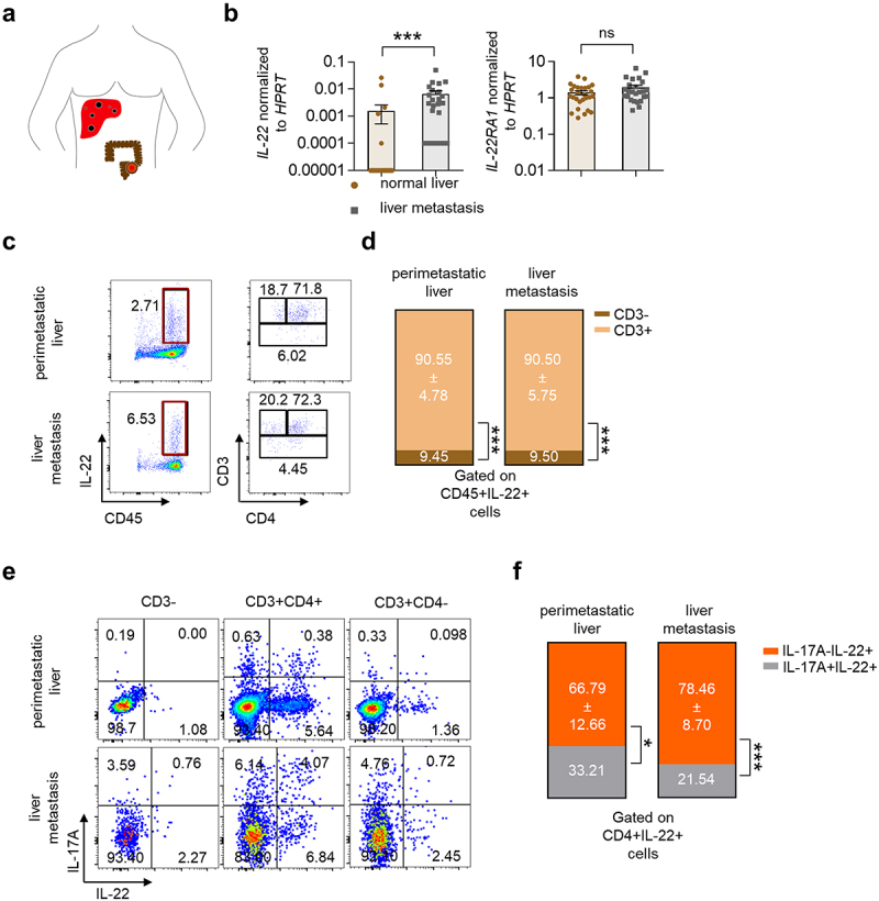
Figure 1.

Increased IL-22 levels in human liver metastasis.
(a) Schematic overview of human liver metastasis. (b) IL22 and IL22RA1 mRNA levels in total liver were measured by qPCR. n ≥ 25 patients per group. (c) Flow cytometry of peri-metastatic and metastatic human liver. (d) Diagram showing the proportion of CD45+IL-22+ cells in peri-metastatic and metastatic human liver. (e) Cells were isolated from fresh peri-metastatic and metastatic liver tissue and analyzed by flow cytometry. (f) Diagram showing the proportion of CD4+IL-22+ cells in peri-metastatic and metastatic human liver. n = 7-10 patients per group. See also Figure S1. Data presented as mean ± SEM. ns > 0.05; ***:p ≤ 0.001 as assessed by the Mann-Whitney U test.
