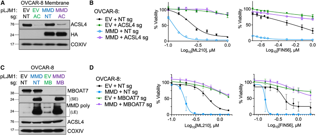
Figure 4. MMD increases ferroptosis susceptibility in an ACSL4- and MBOAT7-dependent manner.
See also Figure S4.
(A) Immunoblot of membrane proteins in OVCAR-8 EV cells transduced with a CRISPR vector containing NT sg (EV + NT sg) or ACSL4 sg (EV + ACSL4 sg) and inOVCAR-8 MMD OE cells transduced with a CRISPR vector containing NT sg (MMD + NT sg) or ACSL4 sg (MMD + ACSL4 sg). Overexpressed MMD was HA tagged. COXIV was used as a loading control.
(B) Viability of cell lines from (A) in response to indicated concentrations of ferroptosis inducers.
(C) Immunoblot in OVCAR-8 EV cells transduced with a CRISPR vector containing NT sg (EV + NT sg) or MBOAT7 sg (EV + MBOAT7 sg) and in OVCAR-8 MMD OE cells transduced with a CRISPR vector containing NT sg (MMD + NT sg) or MBOAT7 sg (MMD + MBOAT7 sg). Overexpressed MMD was HA tagged. The MMD polyclonal antibody was used and images are displayed with both short exposure (SE) and long exposure (LE) times. COXIV was used as a loading control.
(D) Viability of cell lines from (C) in response to indicated concentrations of ferroptosis inducers.
Data points plotted are mean ± SD of n = 3 biological replicates, and experimental figure panels are representative of three independent experiments.

