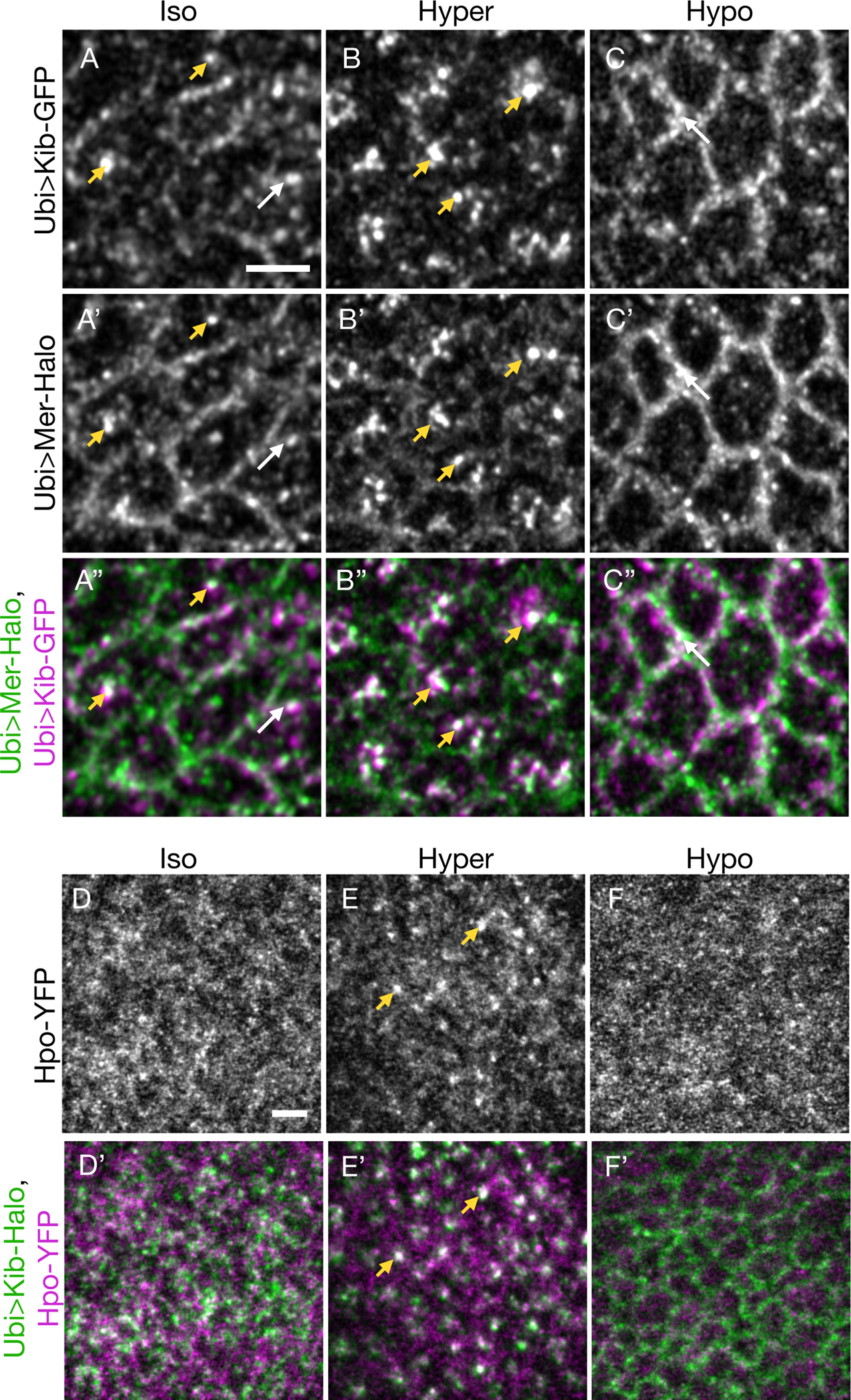
Figure 4. Actomyosin-driven medial Kib assembles a Hippo signaling complex.

(A-C”) Mer displays some co-localization with Kib at the junctional (white arrows) and medial (yellow arrowheads) cortex under isotonic conditions (A-A”). Under hypertonic conditions, Mer is strongly recruited to medial foci with Kib (B-B”). Conversely, under hypotonic conditions, both Mer and Kib localize mainly at the junctional cortex (C-C”).
(D-F’) Hpo is normally diffuse under isotonic conditions (D and D’). However, under the hypertonic shift, Hpo is recruited to the medial cortex with Kib (E and E’). In contrast, Hpo remains diffuse under hypotonic conditions (F and F’). Scale bars = 3μm.
