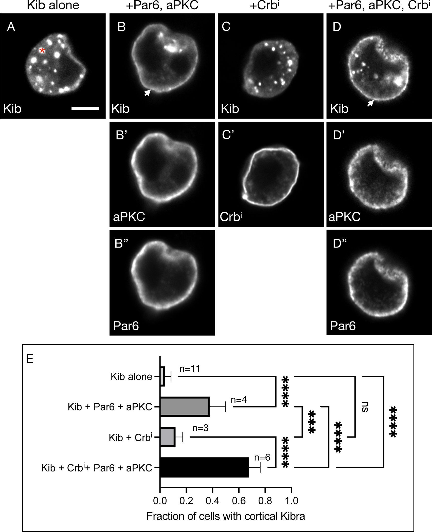
Figure 5. aPKC tethers Kib at the cell cortex in S2 cells.

(A) Expressed by itself, Kib often aggregates in cytoplasmic foci (asterisk) in cultured S2 cells. Scale bar = 5μm.
(B-B”) Co-expression of aPKC and Par6 leads to cortical Kib recruitment (arrowhead in B).
(C-C’) Expression of Crbi alone does not result in cortical Kib recruitment.
(D-D”) Addition of Crbi enhances Par6/aPKC-mediated cortical Kib recruitment.
(E) Quantification of the fraction of cells displaying cortical Kib under the conditions shown in (A-D”). Statistical significance was calculated using One-way ANOVA followed by Tukey’s HSD test; n = number of biological replicates (≥ 50 cells counted per replicate). See also Figure S4.
