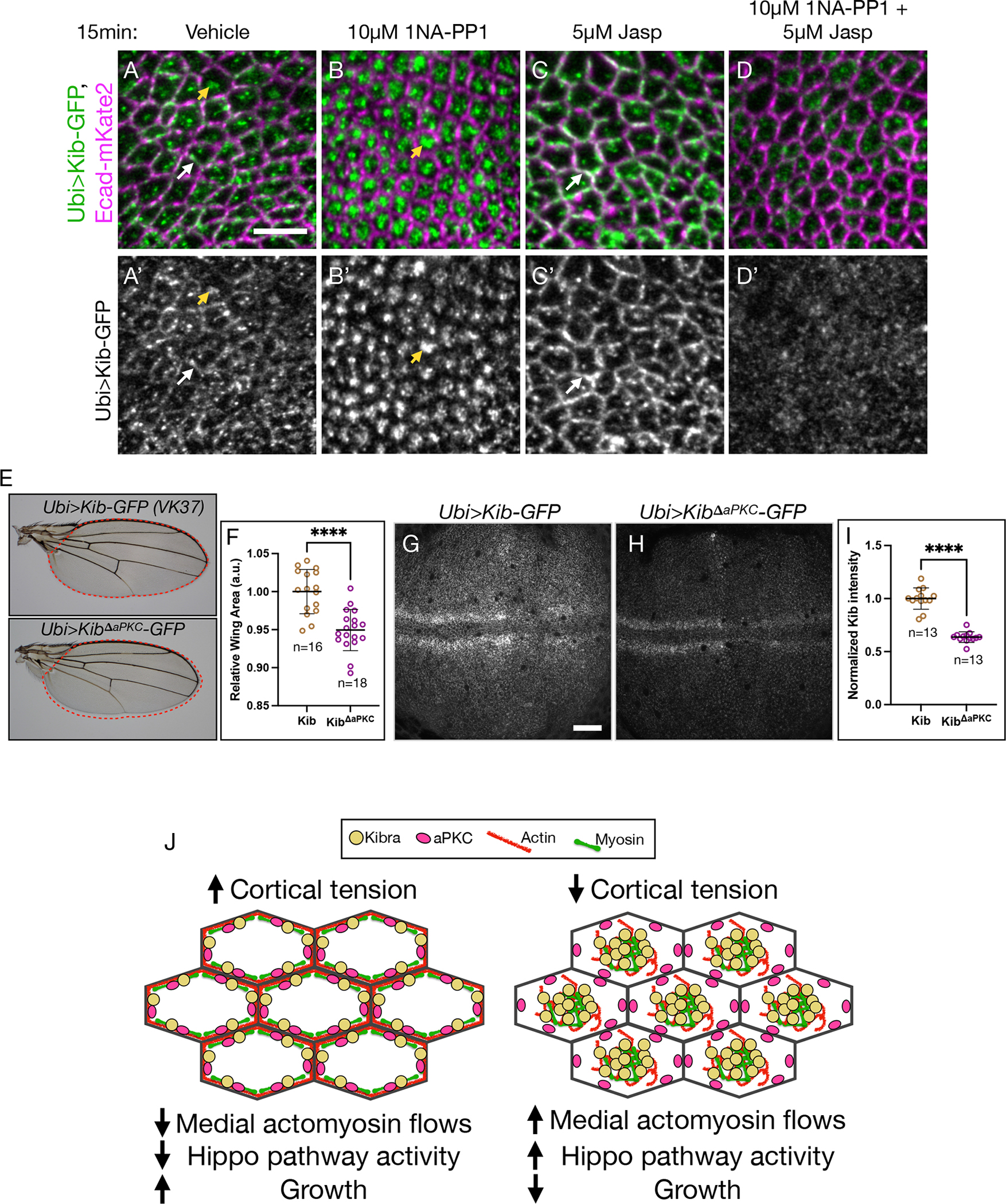
Figure 7. Tethering by aPKC limits Kib-mediated Hippo apathway activation.

(A-D’) In tissues homozygous for aPKCas4 allele, Kib localizes at the junctional (white arrows) and medial (yellow arrowheads) cortex under control conditions (A and A’) but is predominantly medial under aPKC inhibition with 1NA-PP1 (B and B’). Treatment with Jasp leads to more junctional Kib accumulation (C and C’). Under simultaneous treatment with 1NA-PP1 and Jasp, Kib fails to accumulate medially or junctionally (D and D’). Scale bar = 5μm.
(E and F) Wings ectopically expressing KibΔaPKC (Ubi>KibΔaPKC-GFP) are slightly smaller the ones expressing wild-type Kib (Ubi>Kib-GFP); n = number of wings.
(G-I) Wild-type Kib (G and I) is more stable than KibΔaPKC (H and I); n = number of wing dics. Scale bar = 20μm. Transgenes in (E-H) are identically expressed.
Statistical significance in F and I was calculated using Mann-Whitney test.
(J) A cartoon model of Kib regulation via apical polarity and actomyosin flows. Under high cortical tension medial actomyosin flow decreases, allowing aPKC to tether and inhibit Kib at the junctional cortex, resulting in more growth. Under low cortical tension, stronger medial actomyosin flows untether Kib from the junctional cortex and accumulate it medially, where it recruits associated components to promote Hippo signaling and inhibit growth. See also Figure S5 and Figure S6.
