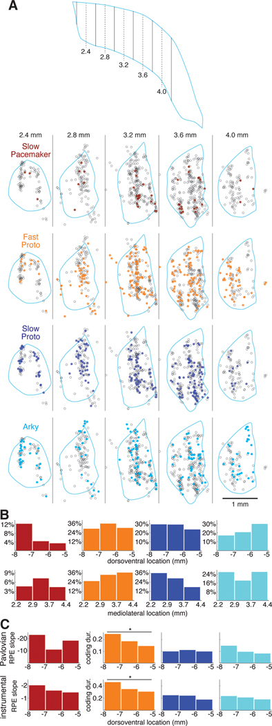
Figure 6. Functional anatomy of GPe subpopulations and value coding.
(A) Locations of each GPe cell recorded in 3 wildtype rats. Top, schematic horizontal section through the GPe illustrating the positions of the virtual sagittal sections used to illustrate cell position below. Dashed lines show the center of each section, solid lines are section boundaries. The far caudolateral tail of the GPe was not sampled. Bottom, each column represents a 400-μm thick sagittal section through the GPe; rostral is to the left and dorsal is up. Each circle represents the location of one recorded neuron within that section; the locations of individual cells recorded at the same site have been jittered slightly so that they can be distinguished (see Methods). Each section is overlaid with the outline (pale blue) of the GPe of one of the rats from a location in the mediolateral center of the section. Some cells appear outside of this outline because the GPe shifts significantly within each section (e.g., the GPe curves caudally as it extends laterally) and due to variation across rats, but all were verified to be within the GPe. Each row features one GPe cell type; cells of that type are filled circles while the other cells are open circles. See also Figure S6.
(B) Location distributions by cell type. The GPe was divided into 3 sectors along the dorsoventral (top) or mediolateral (bottom) axis. The height of each bar gives the percentage of cells within that sector that are members of a given cell type.
(C) Value coding by dorsoventral location. The height of each bar gives the mean regression slope following reward (Slow Pacemakers) or the fraction of time spent coding value (all other cell types) in each dorsoventral sector. Top row, Pavlovian conditioning; bottom row, instrumental learning. Y-axis scale in the left column (regression slope in Slow Pacemakers) is inverted.

