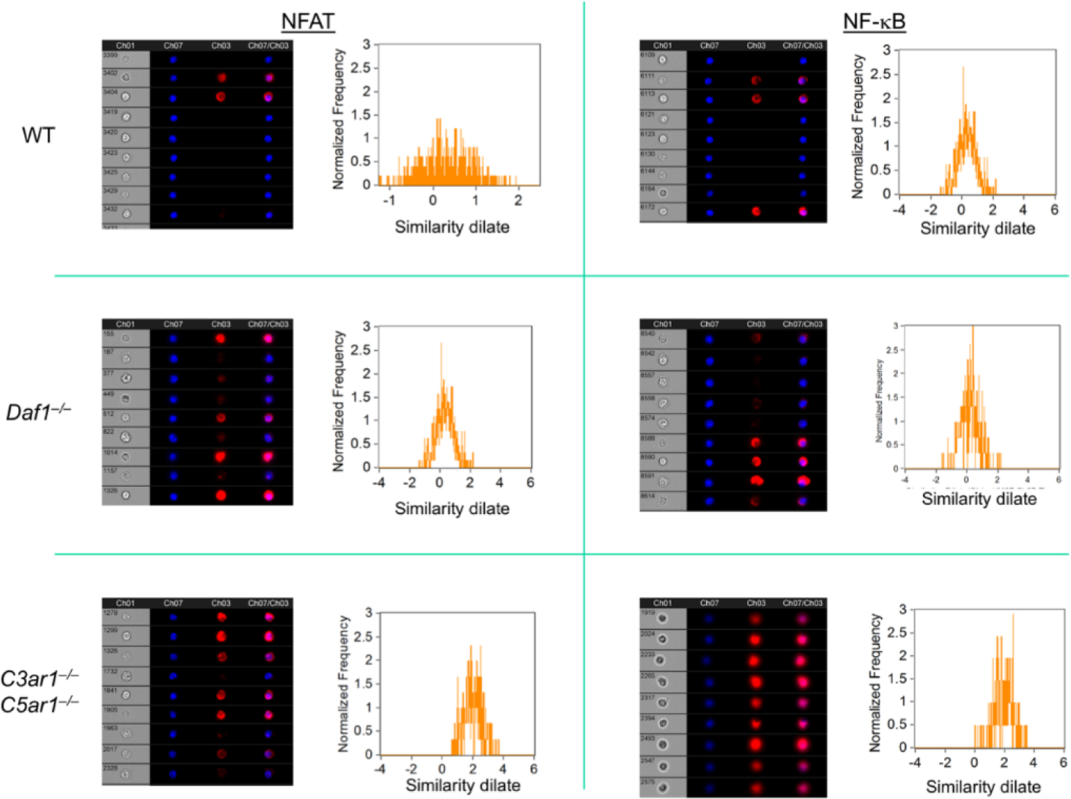
Figure 2: Nuclear translocation of NFAT and NF-kB in Tregs devoid of C3ar1/C5ar1 signaling.

Sorted induced Foxp3+ cells on each genotype were assayed for nuclear NFAT and NF-kB by Amnis Cytometry. The flow plots show colocalization of NFAT and NF-kB with the nuclear stain. Representative of 2 assays. The red lines are the anti-TGF-β and anti-IL-10 stains. The black lines are nonrelevant controls.
