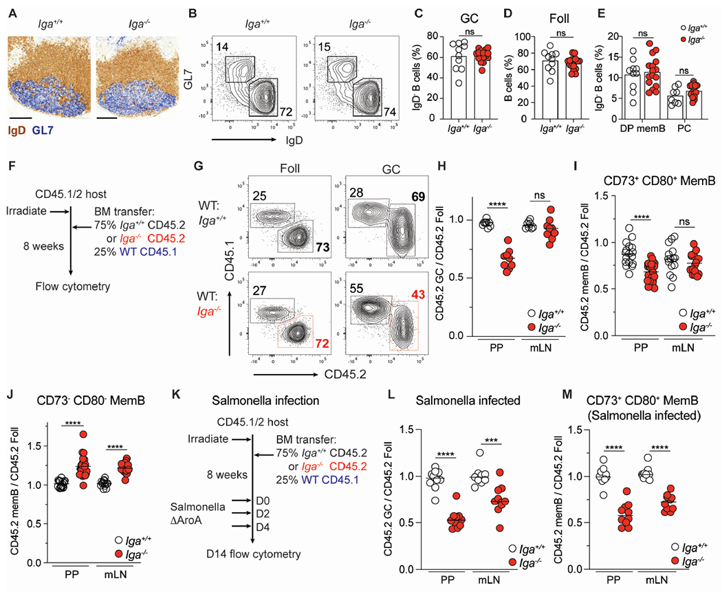
Figure 1. IgA+ B cells dominate the germinal center reaction and are preferentially selected into the memory compartment.

(A-E) Steady state, co-housed, 8–12-week-old Iga+/+ and Iga−/− mice.
(A) Frozen PP sections from Iga+/+ and Iga−/− mice stained with immunohistochemistry antibodies against IgD (brown) and GL7 (blue). Scale bars, 200 μm.
(B) Representative plot of PP GC (GL7+ IgD−) and Foll (GL7− IgD+) B cells in Iga+/+ and Iga−/− mice. Gated on live CD138− B cells.
(C) PP GC B cells as percentage of activated (IgD−) B cells.
(D) PP Foll B cells as percentage of B cells.
(E) CD73+ CD80+ (DP) MemB, PCs as percentage of activated B cells.
(F) Iga−/− mixed BM chimera experimental set up for (G-J).
(G) Representative CD45 staining on Foll and GC B cells in mixed BM chimeras.
(H) Ratio of frequency of CD45.2 GC B cells to CD45.2 Foll B cells in PP and mLN.
(I, J) Ratio of frequency of CD45.2 DP memB cells (I) or CD45.2 CD73− CD80− (DN) MemB cells (J) to CD45.2 Foll B cells in PP and mLN.
(K) Salmonella infection experimental setup in Iga−/− mixed BM chimera for (L-M).
(L) Ratio of frequency of CD45.2 GC B cells to CD45.2 Foll B cells in PP and mLN of Salmonella infected chimeras.
(M) Ratio of frequency of CD45.2 DP MemB cells to CD45.2 Foll B cells in PP and mLN of Salmonella infected chimeras.
Data from at least 3 independent experiments with 2-3 mice per group (A-M). Each symbol represents one mouse. ns=not significant; ***p < 0.0005; ****p < 0.0001; Unpaired two-tailed Student’s t-test.
See also Figure S1.
