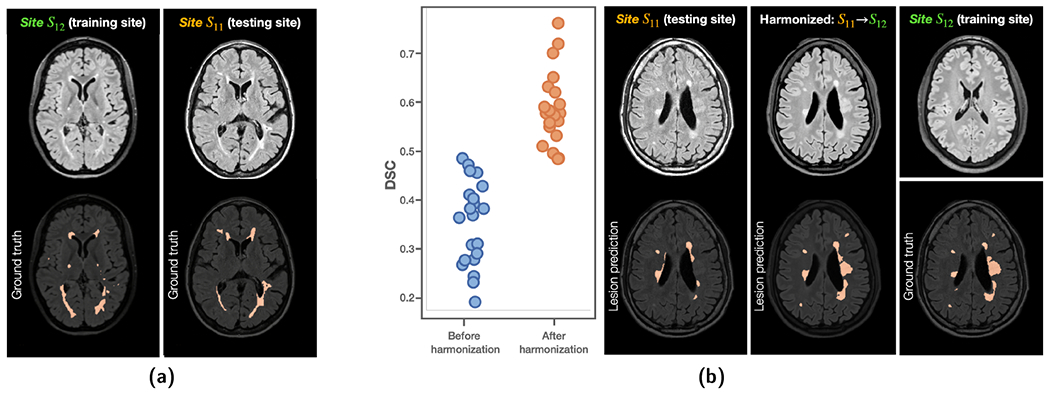
Figure 12:

(a) Training and testing sites for WM lesion segmentation with a 3D U-Net. (b) DSC showed improvements after harmonizing images from the testing site (Site S11) to the lesion training site (Site S12). Example images are shown on the right.
