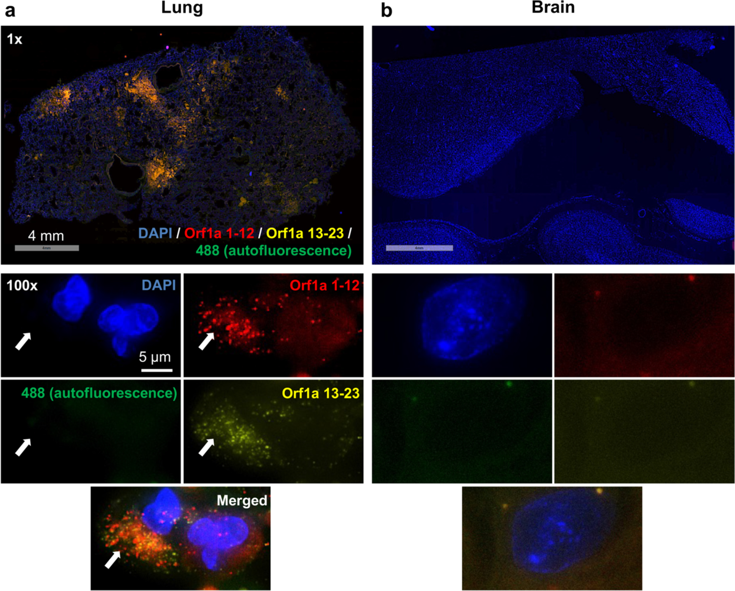
Fig. 1. No SARS-CoV-2 viral RNA detectable in the brain tissue.

(a) The presence of SARS-CoV-2 viral RNA in the lung tissue, potentially associated with epithelial cell infection, was revealed through hybridization with probes of SARS-CoV-2 Orf1a (coupled with amplifier labeling Alexa Fluor 647 to Orf1a 1–12 and Alexa Fluor 546 to Orf1a 13–23). Amplifier labeling Alexa Fluor 488 green was used as the negative control marking tissue autofluorescence to minimize false positivity. (b) In contrast, no SARS-CoV-2 viral RNA was detected in the brain tissue. Scale bars 4 mm for the top low power images, 5 μm for the below high power images.
