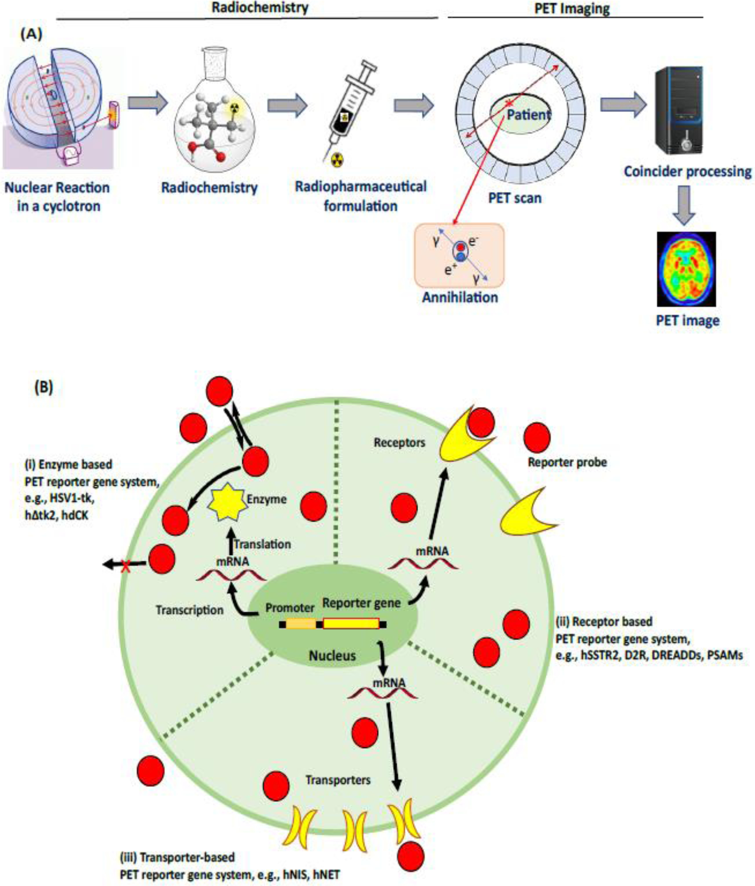
Figure 1: PET imaging: an overview.

A) Schematic representation of brain imaging with PET. A cyclotron generates radionuclides which are used for the production of PET reporter probes via one or more chemical reactions (radiochemistry). A final radiopharmaceutical formulation is prepared with a suitable vehicle after the isolation of the PET reporter probe using HPLC, and then injected into living subjects. PET imaging involves the reconstruction of the location of reporter probe within the body, via data acquisition and post-processing (see Text box 1 for details). B) Overview of the expression of reporter gene systems; three different mechanisms by which PET radiotracers image reporter gene expression based on (i) enzymes: intracellular entrapment of radiotracers after phosphorylation by enzymes encoded by reporter genes, e.g. HSV1-tk phosphorylates pyrimidine-based radiotracers, such as [18F]FIAU, trapping the phospohorylated ligand inside the cell (ii) receptors: radiotracers bind protein receptors encoded by reporter genes, e.g. the human somatostatin receptor type 2 (hSSTR2), dopamine D2 receptor (D2R), and optogenetic-based receptors, encoded by a reporter gene (iii) transporters: accumulation of radiotracers into cells mediated by membrane transporters encoded by reporter genes, e.g. the human sodium–iodide symporter (hNIS), and human norepinephrine transporter (hNET) encoded by reporter gene.
