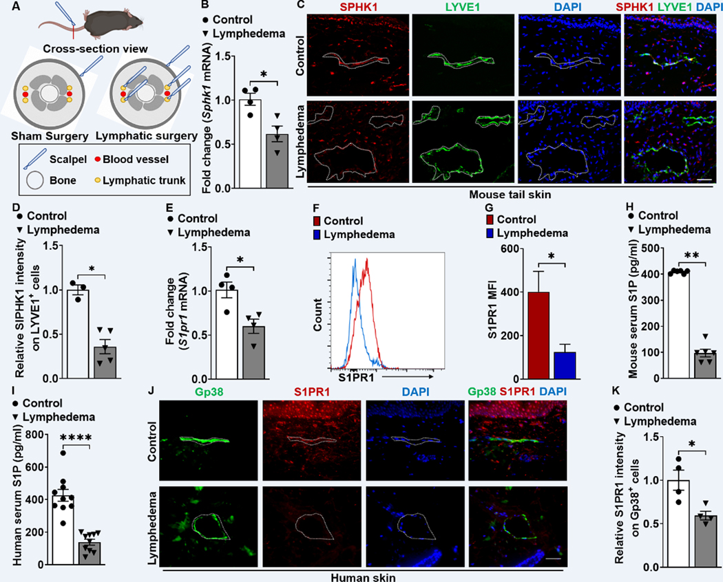
Figure 1. S1PR1 signaling of LECs is reduced in both mouse and human lymphedema skin.
(A) Acquired lymphedema was surgically induced in the tails of C57BL/6J mice through thermal ablation of lymphatic trunks. Skin incision, alone, was performed in sham surgery groups. (B) RT-qPCR analysis of Sphk1 mRNA levels in tail skin from control sham surgery mice or animals subjected to lymphatic surgery (n = 4 per each group). (C) Representative immunofluorescence (IF) staining of SPHK1 (red) and LYVE1 (green) of the skin tissues harvested from control or lymphedema mice. DAPI (blue) stains for the nucleus. Scale bar = 40 μm. (D) Quantification of the SPHK1 staining intensity comparing groups shown in C (n ≥ 3 per each group). (E) RT-qPCR analysis of S1pr1 mRNA levels in tail skin from control sham surgery mice or lymphedema mice (n = 4 per each group). (F and G) Flow cytometry histograms show the mean fluorescence intensity of S1PR1 on LEC (Gp38+CD31+) population. Representative (F) and compiling data (G) are shown (n ≥ 3 per each group). (H and I) The serum concentration of S1P in mouse (H) and human (I) lymphedema (n = 10 per each group). (J) Representative IF staining of Gp38 (green) and S1PR1 (red) of the human skin from healthy control or lymphedema. DAPI (blue) stains for the nucleus. Scale bar = 50 μm. (K) Quantification of the S1PR1 intensity comparing groups shown in J (n = 4 per each group). Data are presented as mean ± SEM; * p < 0.05, ** p < 0.01, and **** p < 0.0001 by the Mann-Whitney test.

