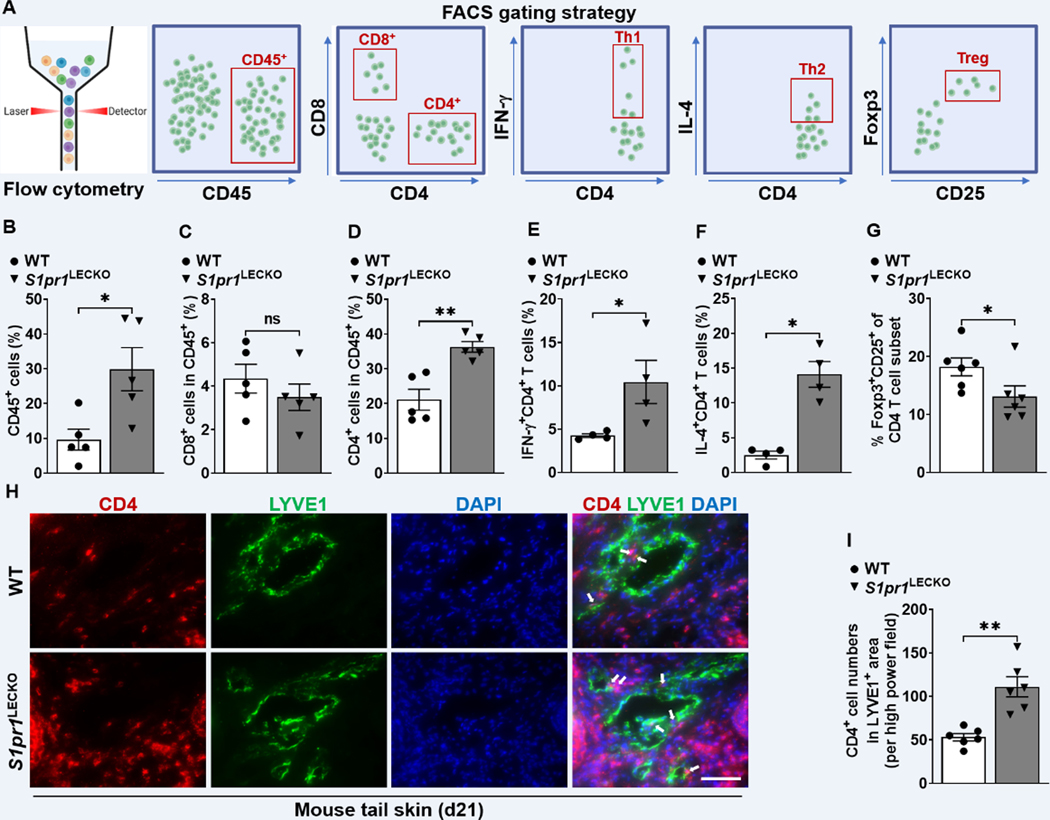
Figure 3. LEC S1pr1 deficiency promotes CD4 T cell infiltration following lymphatic surgery.
(A) Flow cytometric gating scheme for determining tail skin immune cell populations. Flow cytometric analysis was performed d21 after lymphatic surgery. (B-G) Quantification of CD45+ cells (B), CD8+ T cells, (C), CD4+ T cells (D), CD4+IFN-ɣ+ Th1 cells (E), CD4+IL-4+ Th2 cells (F), and Foxp3+CD25+CD4+ Treg cells (G) in tail skin (n ≥ 4 per each group). (H) Representative IF staining of CD4 (red) and LYVE1 (green) of the lymphedema mouse tail skin from WT or S1pr1LECKO mice. DAPI (blue) stains for the nucleus. Arrows indicate CD4 T cells surrounding lymphatic vessels. Scale bar = 50 μm. (I) Quantification of the CD4 T cell staining presented in H (n ≥ 5 per each group). Data are presented as mean ± SEM; * p < 0.05, ** p < 0.01, and ns (not significant) by the Mann-Whitney test.

