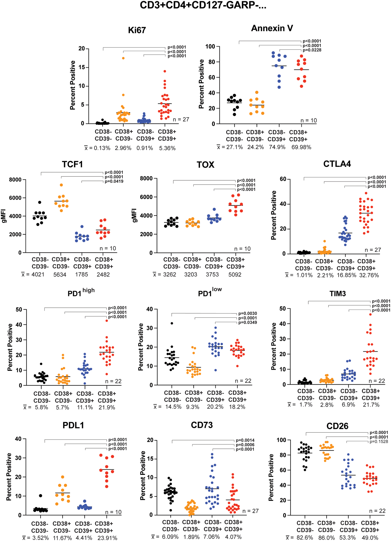
Figure 2. Teee have increased expression of proliferation, apoptosis, exhaustion, and co-inhibitory markers.

Baseline peripheral blood cells from melanoma patients were stained and analyzed by flow cytometry. The CD3+CD4+CD127-GARP- parent population was gated and the CD38/CD39 quadrants assessed for expression of the indicated markers. The CD38-CD39- population is shown in black, CD38+CD39- in orange, CD38-CD39+ in blue, and CD38+CD39+ (Teee) in red. Corresponding sample sizes assessed are indicated in the lower right of each panel. Population sample mean values are given below each group and represented by horizontal bars in the graphs. Significance was determined by repeated measure ANOVAs with Dunnett’s multiple comparison tests of the Teee population against the other CD38/CD39 quadrants.
