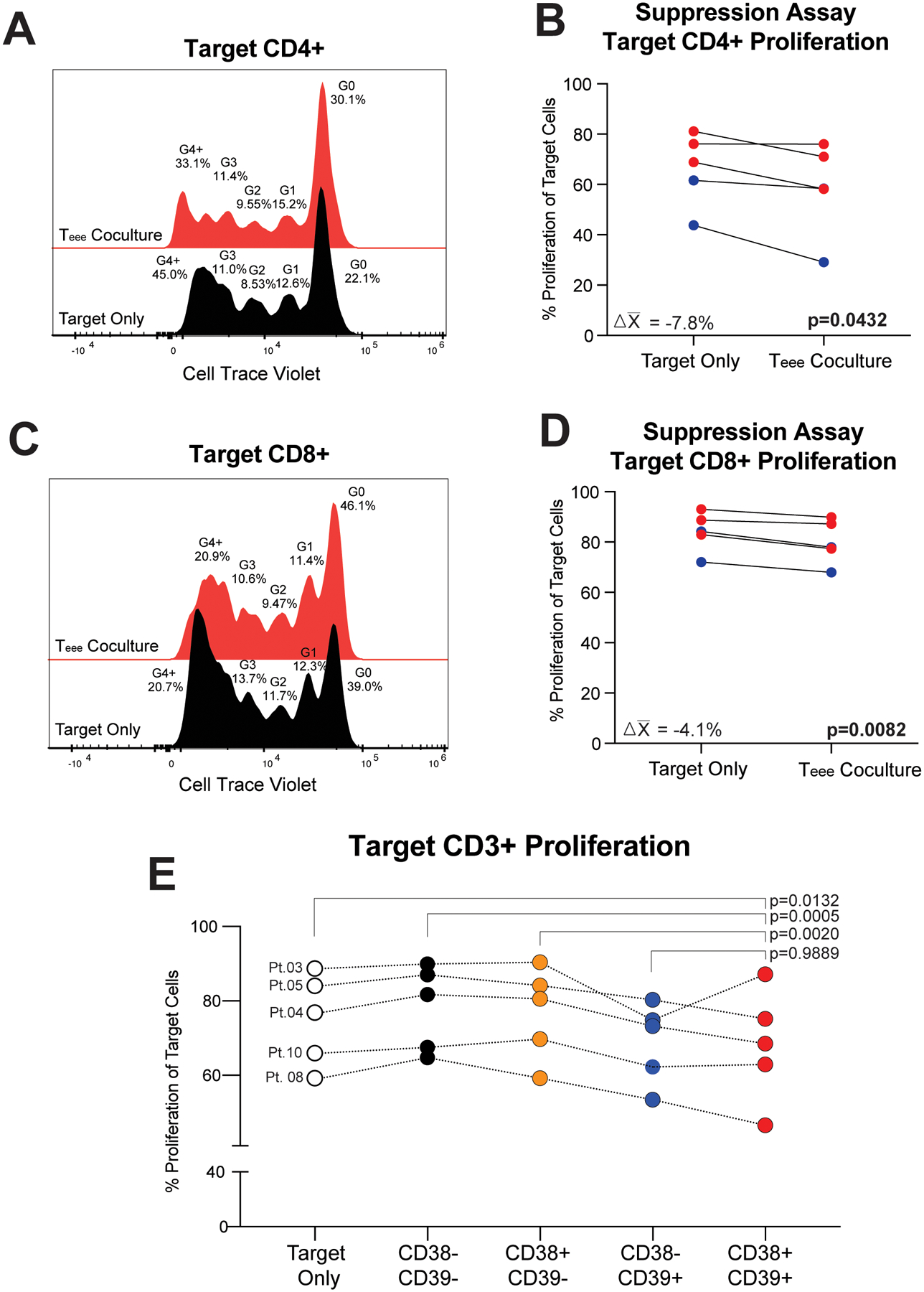
Figure 5. Teee suppress proliferation of autologous T-cells in vitro.

Baseline peripheral blood cells from metastatic melanoma patients were flow sorted for Teee. Autologous bulk CD3+ T-cells as targets were stained with Cell Trace Violet, activated by plate bound αCD3 and soluble αCD28, and cultured alone or in the presence of Teee at a target to Teee ratio of 2:1. (A) A representative plot of Cell Trace Violet is shown for target CD4+ T-cells alone (black histogram) or in the presence of Teee (red histogram). (B) The percentages of proliferating target CD4+ T-cells for five samples evaluated are shown. Lines connect paired patient samples. P-values were determined by paired t-tests. Mean changes in the percentage of proliferating target cells are given in the corresponding panel. Red coloring indicates specimens from non-responding patients and blue coloring represents responding patients. (C,D) Target CD8+ T-cells are likewise shown. (E) Target CD3+ T-cells were cultured alone or in the presence of autologous CD3+CD4+CD127-GARP-CD38-CD39- (black dots), CD3+CD4+CD127-GARP-CD38+CD39- (orange dots), CD3+CD4+CD127-GARP-CD38-CD39+ (blue dots), or CD3+CD4+CD127-GARP-CD38+CD39+ (Teee, red dots). The percentages of proliferating target CD3+ T-cells for five samples evaluated are shown. Lines connect paired patient samples. P-values were determined by repeated measures ANOVA and Dunnett’s posthoc tests.
