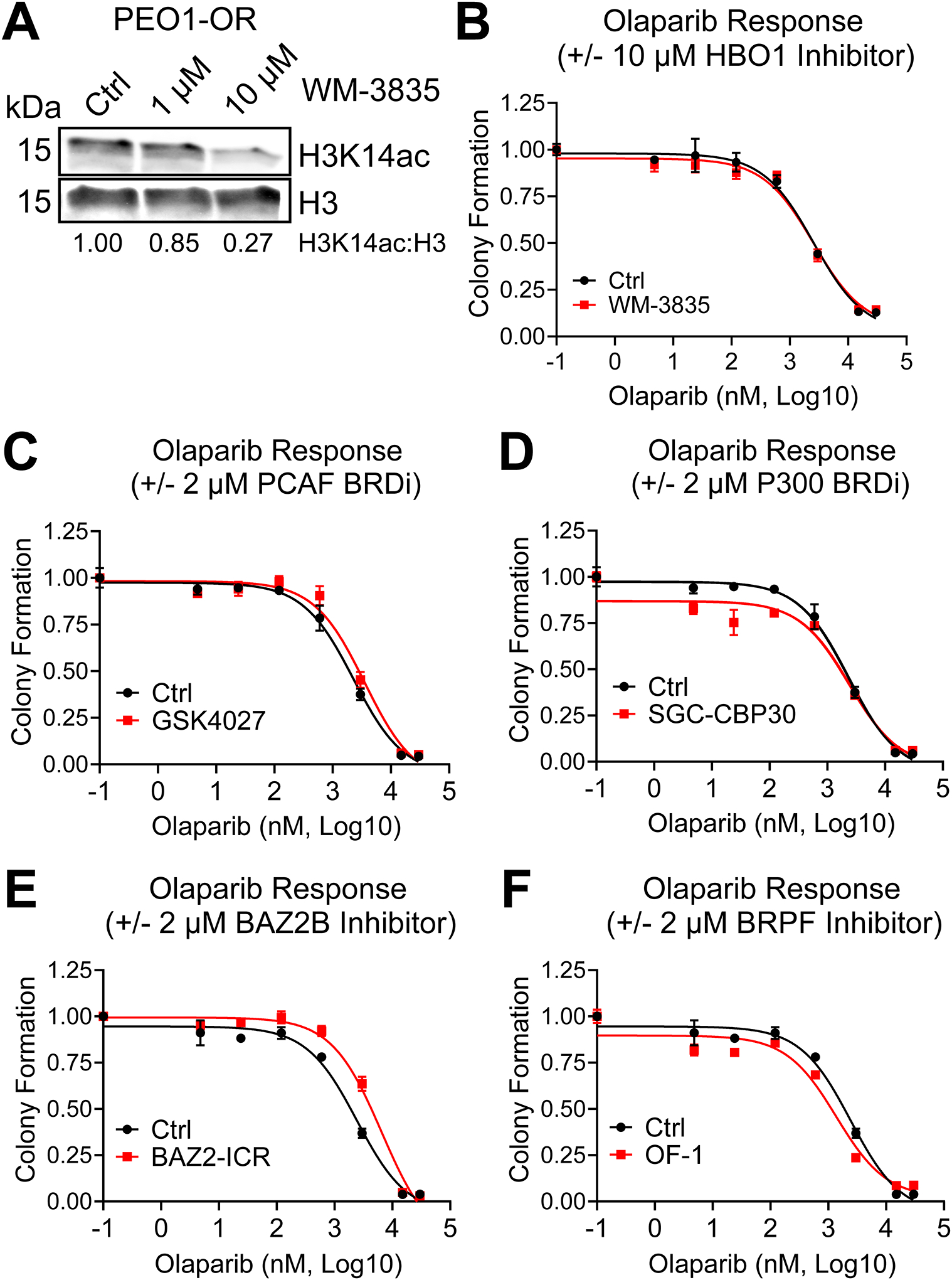
FIG 3. Depletion of H3K14ac does not shift olaparib IC50, but specific BRD inhibitors moderately affect olaparib response in PEO1-OR HGSOC cells.

(A) PEO1-OR cells were treated with the indicated doses of HBO1 acetyltransferase inhibitor WM-3835 for 6 hours. Histone extracts were analyzed by immunoblot for H3K14ac and total H3. The H3K14ac:H3 ratio was calculated by densitometry to quantify H3K14ac depletion. (B) PEO1-OR were seeded at 2500 cells per well in a 24-well plate and treated with increasing doses of olaparib with or without 10 μM WM-3835. Media and drug were changed every 2–3 days for eight days, after which colonies were fixed and stained with crystal violet. Stain was dissolved and the absorbance from each well was read by spectrophotometer, then normalized to vehicle control. Error bars, SEM. Olaparib IC50 for each treatment condition was calculated in GraphPad Prism. (C-F) PEO1-OR cells in a 24-well plate were treated with increasing doses of olaparib with or without 2 μM of the indicated BRD inhibitors. Colony formation and olaparib IC50 were determined as described in Fig. 3B. Error bars, SEM.
