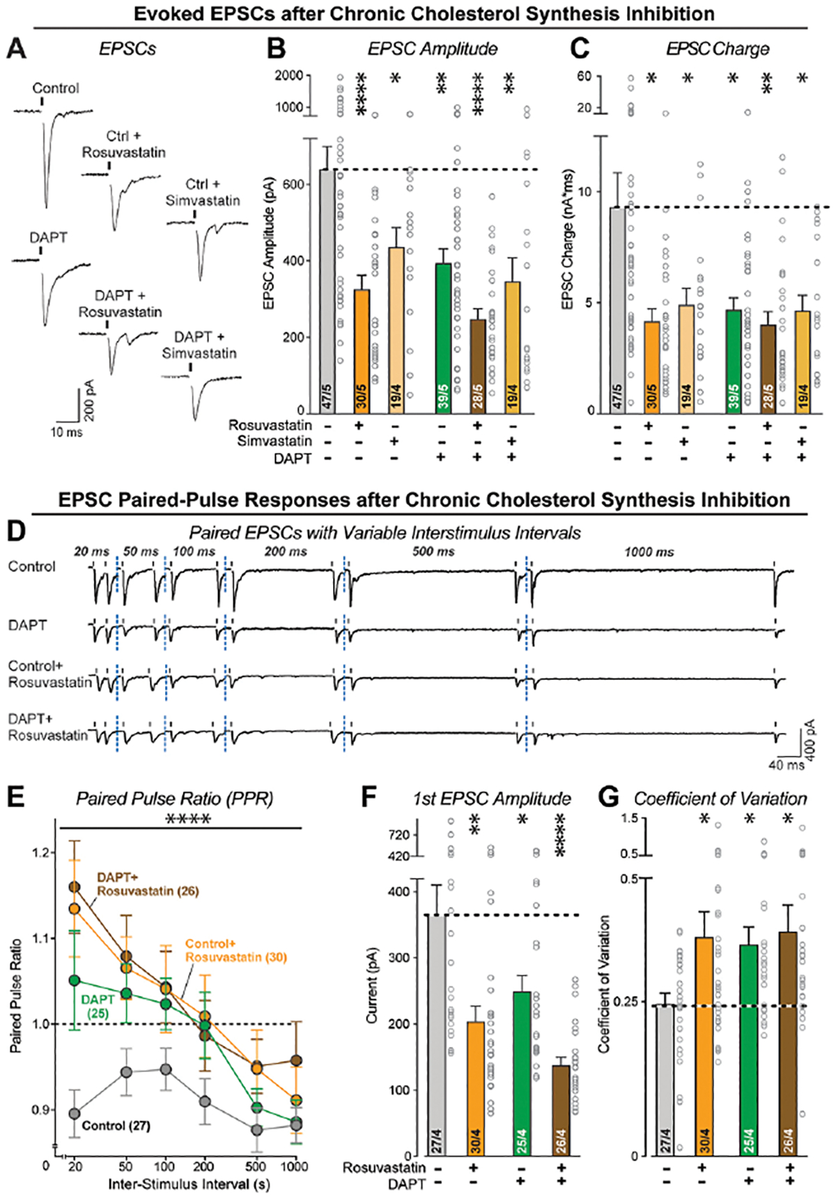
Figure 8: HMG-CoA reductase inhibitors (statins) suppress the neurotransmitter release probability in human neurons.

(A) Representative EPSC traces from neurons treated with DAPT and HMG-CoA reductase inhibitors as described for A & B.
(B & C) Chronic inhibition of HMG-CoA reductase causes the same suppression in synaptic strength, measured via the amplitude (B) or charge transfer (C) of evoked EPSCs, as chronic inhibition of γ-secretase. Note that the effects of chronic inhibition of HMG-CoA reductase of γ-secretase are not additive.
(D) Representative traces of EPSCs evoked by two closely spaced stimuli (paired-pulse stimulation), monitored in human neurons as a function of chronic treatments with γ-secretase and/or HMG-CoA reductase inhibitors.
(E) Chronic suppression of the HMG-CoA reductase and/or γ-secretase activity reverses paired-pulse depression, suggesting a decline in release probability. The summary plot depicts the paired-pulse ratios (second/first EPSC amplitude) as a function of the interstimulus interval.
(F) As shown for the isolated EPSCs, chronic suppression of the HMG-CoA reductase and/or γ-secretase activity decreases the amplitude of evoked EPSCs monitored via the first response during paired-pulse stimulation.
(G) Chronic suppression of the HMG-CoA reductase and/or γ-secretase activity also increases the coefficient of variation of evoked EPSC amplitudes, consistent with a reduced release probability.
Data are means +/− SEM (the number of cells/biological replicates are shown in the bars). For (B), (C), (F), (G), statistical analysis was measured using two-tailed unpaired t-test. For (E), statistical analysis was assessed by two-way ANOVA with Bonferroni’s multiple comparison test. Statistical significance: *p<0.05; **p<0.01, ***p<0.001. For additional data, see Figure S8.
