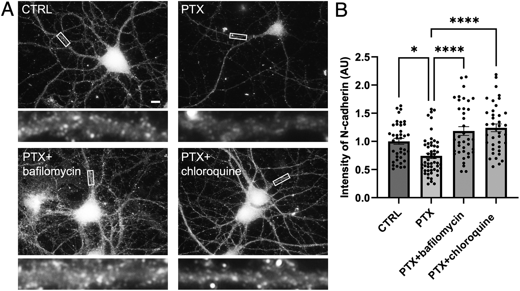
Figure 8. N-cadherin is degraded by the lysosome.

(A) Immunostaining of endogenous N-cadherin in cultured hippocampal neurons left unstimulated (CTRL), treated with PTX alone, or treated with PTX in combination with lysosome inhibitor bafilomycin or chloroquine. Higher magnification views of representative secondary dendrites (boxed regions) are shown below each neuron. Scale, 10 μm. (B) Quantification of images in (A) (p< 0.0001, F=14.87, DF=3) (n=39–52 neurons; one-way ANOVA and Tukey’s multiple comparison test, *p<0.05, ****p<0.0001, ns=nonsignificant).
