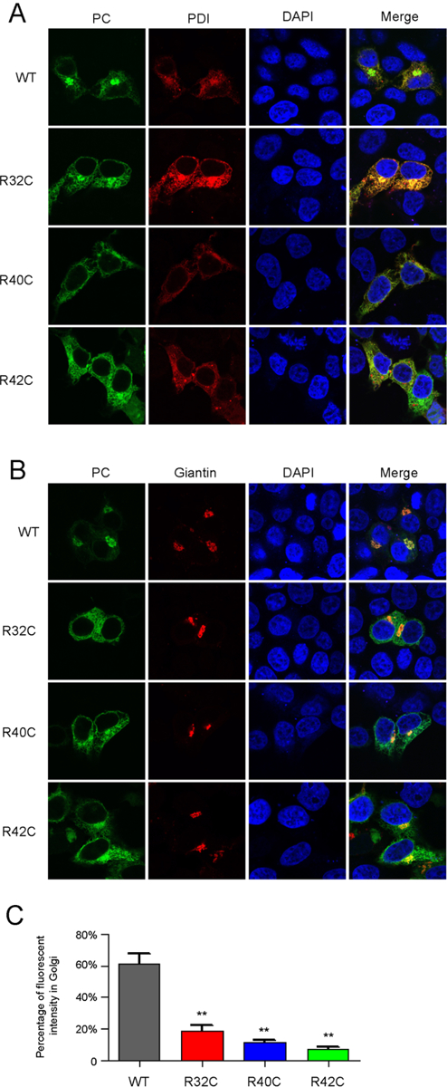
Figure 3. Mutations of R32C, R40C and R42C result in ER retention of the PCspg-EGFP reporter.

A and B. Subcellular localization of the PCspg-EGFP reporter protein in ER (A) or Golgi (B). The PDI-mCherry (A) and mScarlet-Giantin (B) were used as ER marker and Golgi marker, respectively. The nucleus was stained by DAPI. The cells were culture in medium with 10μM vitamin K. C. Quantification of fluorescent intensity. Green fluorescent intensity of the whole cell and Golgi in Figure 3B was quantified by ImageJ. The percentage of reporter’s fluorescent intensity in Golgi was calculated by dividing the fluorescent intensity in Golgi to that in the whole cell. Eight to ten cells were analyzed for each construct, and the t-test was performed between wild-type and mutants by Graphpad Prism 7. ** indicates p<0.01.
