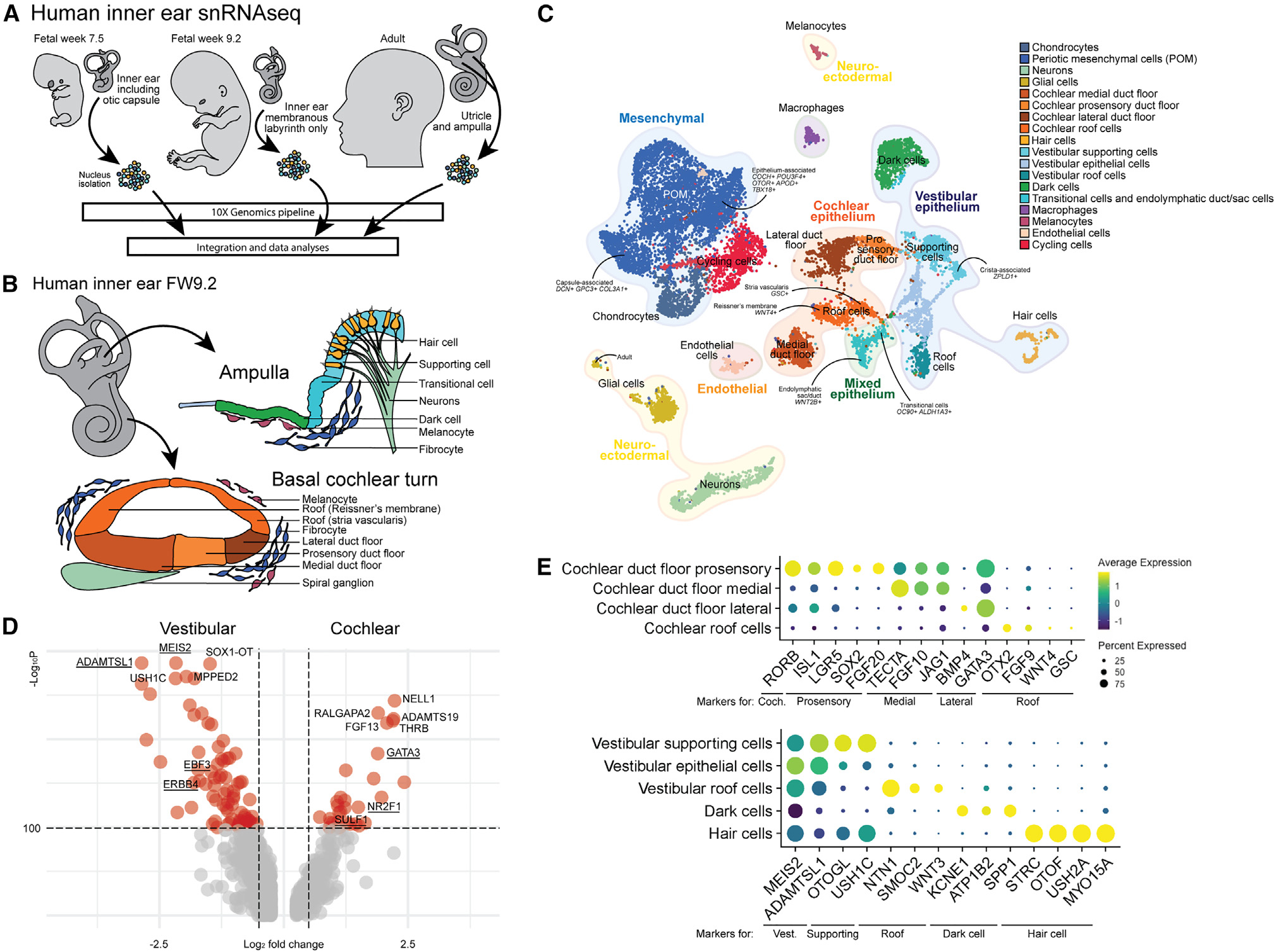
Figure 3. Generation of the human fetal and adult inner ear single-cell atlas.

(A) Experimental overview of fresh human inner ear tissue collection from FW7.5 (n = 1), FW9.2 (n = 1), and adult donor (n = 1).
(B) The human inner ear at FW9.2, illustrating cellular diversity in the vestibular ampulla and basal turn of the cochlea.
(C) UMAP plot of combined human inner ear datasets with cell type annotations.
(D) Volcano plot showing differentially expressed genes between vestibular supporting cells and cochlear duct floor. Underlined genes are described to be differentially expressed between vestibular and cochlear cell types.
(E) Dot plot displaying standardized expression of known marker genes in cochlear and vestibular cell types.
Coch., cochlear; Vest., vestibular. See also Figure S6.
