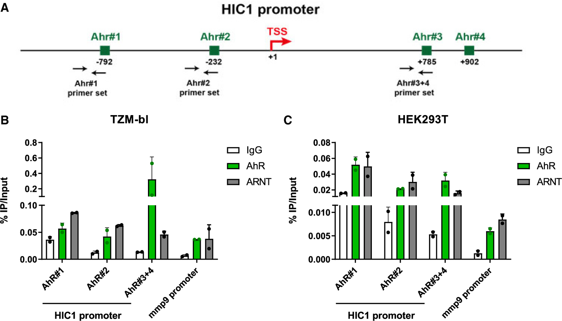
Figure 7. The AhR/ARNT complex is recruited in vivo to the HIC1 promoter.

(A) Schematic representation of the HIC1 promoter, with the localization of the potential AhR-/ARNT-binding sites identified by in silico analysis (using the Jaspar 2022 consensus motif) and with the localization of the ChIP-qPCR primer sets.
(B and C) Chromatin prepared from TZM-bl (B) or HEK293T (C) cells was immunoprecipitated using specific Abs directed against AhR and ARNT or using an immunoglobulin G (IgG) as background measurement. Results are presented as histograms indicating percentages of immunoprecipitated DNA compared with the input DNA (% IP/input). Data are the means ± SD from one experiment representative of four independent experiments. Shown are bars that indicate mean ± SD values.
