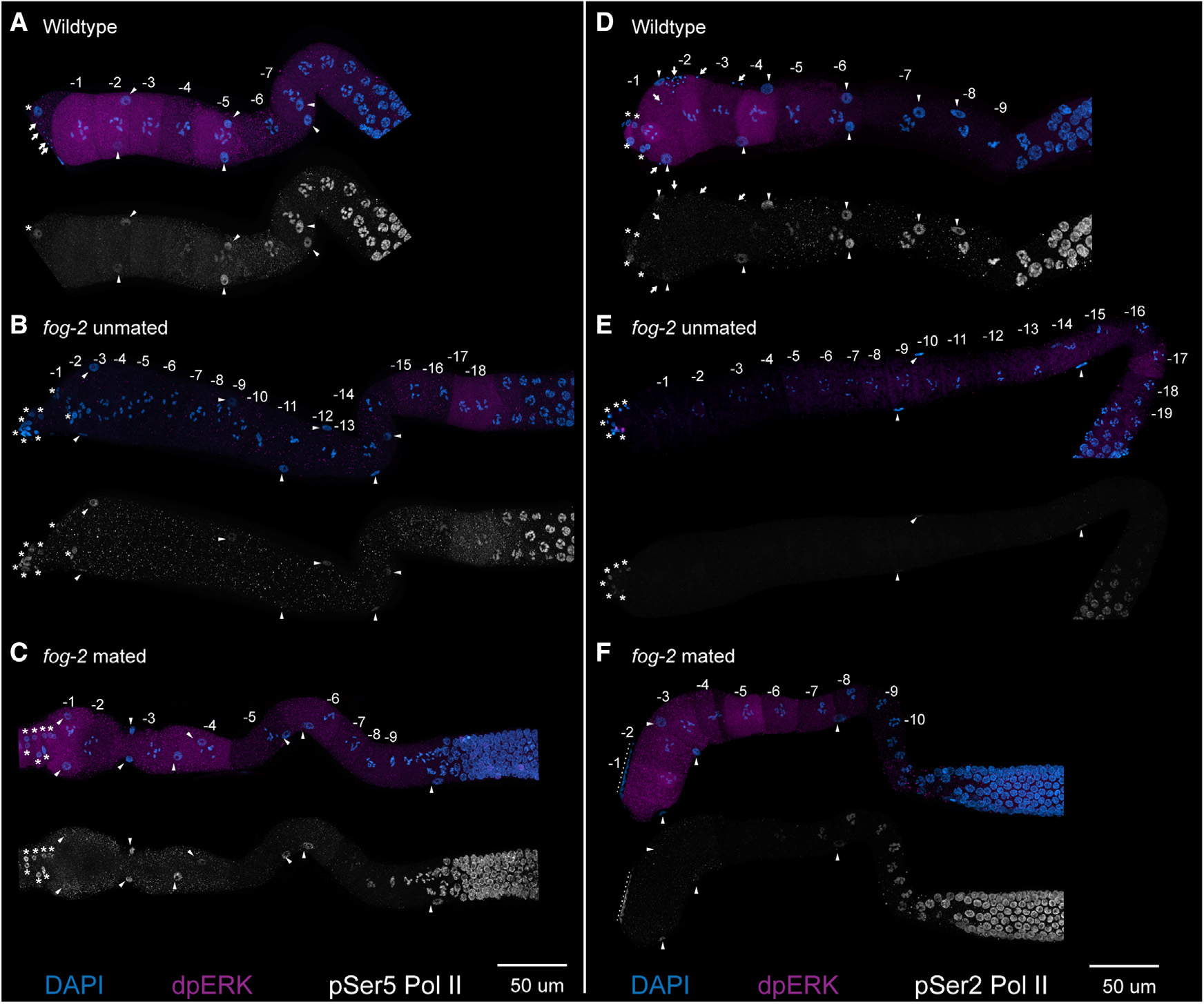
Figure 3. Sperm signal and MPK-1 ERK activation do not induce activation and phosphorylation of RNA Pol II Ser5 or Ser2.

(A–F) Maximum-intensity projections from dissected germlines from (A and D) wild type, (B and E) feminized (fog-2) unmated, and (C and F) fog-2 mated for 2 h are shown with numbered oocytes. Germlines are stained with DAPI (blue), dpMPK-1 (magenta), and either pSer5 Pol II (white, A–C) or pSer2 Pol II (white, D–F). The nuclei of sheath cells (triangles), spermathecal cells (asterisks), and sperm (arrows) are labeled. Nuclei near the dotted lines in (F) are from a neighboring germline. Scale bars, 50 μm.
