
Figure 5. Class I transcripts are present in the arrested −1 oocyte and the spermatheca.
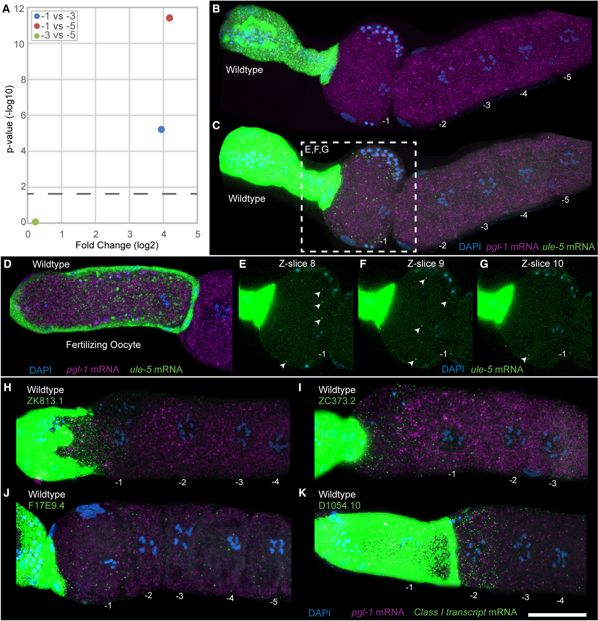
(A) Plot showing the fold change in ule-5 between different oocyte positions. The p value cutoff (horizontal dashed line) was p < 0.05.
(B–G) HCR-FISH staining of dissected wild-type germlines with DAPI (blue), ule-5 mRNA (green), and pgl-1 mRNA (magenta) with oocytes numbered, acquired as a z stack with 14 slices with a step size of 1.5 μm. (B and C) Maximum-intensity projection images through the full z stack with (B) normal acquisition intensity for ule-5 mRNA and (C) enhanced intensity for ule-5 mRNA. (D) Dissected wild-type germline with oocyte in the spermatheca. (E–G) Individual slices from central planes (−1 nucleus visible) of the z stack with enhanced intensity, ule-5 FISH signal (arrowheads) and slice number labeled. For −2, −3, −4, and −5 oocytes and end slices 1 and 14, see Figure S7.
(H–K) Dissected and stained germlines showing representative expression patterns for the class II transcripts (H) ZK813.1, (I) ZC373.2, (J) F17E9.4, and (K) D1054.10. Shown are maximum intensity projection images through the full z stack DNA (blue), pgl-1 mRNA (purple), and target mRNA (green). Scale bar, 25 μm. See also Figures S7, S8, and S9.