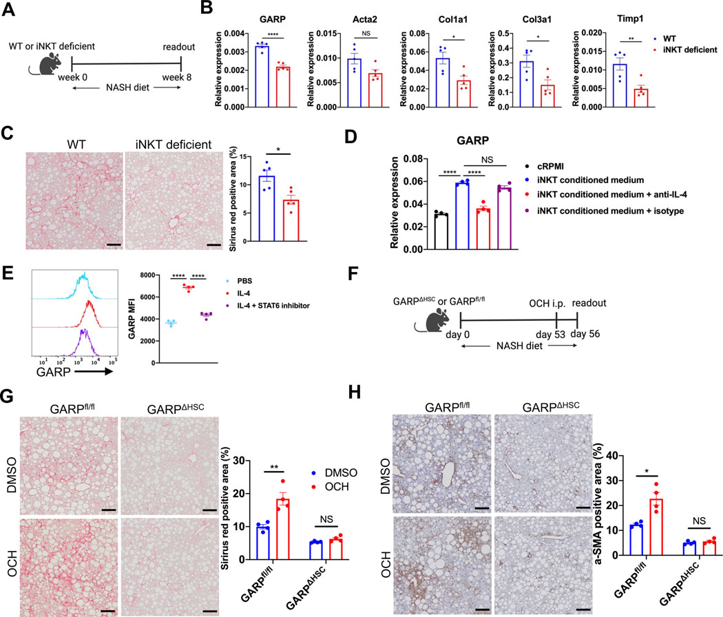
Fig. 3. NKT cells promote fibrotic scarring by inducing GARP expression on HSCs through IL-4 production.
(A) Experimental design of NASH diet-induced liver fibrosis in wild-type (WT) or Traj18-knockout (iNKT-deficient) mice. (B) GARP and fibrotic gene expression in the liver of mice detected by qRT-PCR. (C) Representative images of Sirius red staining of mouse liver (left) and quantification of stained area (right). Scale bar 100 μm. (D) GARP mRNA expression in HSCs treated with complete RPMI (cRPMI) or iNKT-conditioned medium in the presence or absence of anti-IL-4 or isotype control antibody. (E) Representative histogram plot (left) and mean fluorescent intensity (MFI) (right) of GARP staining on the surface of HSCs after incubation with PBS, recombinant IL-4, or IL-4 plus STAT6 inhibitor AS1517499. (F) Scheme of OCH injection during NASH diet-induced liver fibrosis. (G) Representative images of Sirius red staining of mouse liver (left) and quantification of stained area (right). Scale bar 100 μm. (H) Representative images of IHC staining of α-SMA in the liver of mice (left) and quantification of stained area (right). Scale bar 100 μm. Each data point represents a single mouse in A-C and F-H or one well in D-E. Data are presented as mean ± SEM and are from one experiment representative of at least two independent experiments with similar results. Unpaired t-test in B-C, G-H and one-way ANOVA in D-E were used to compare two groups. *p<0.05;**p<0.01;***p<0.001;****p<0.0001; NS, not significant.

