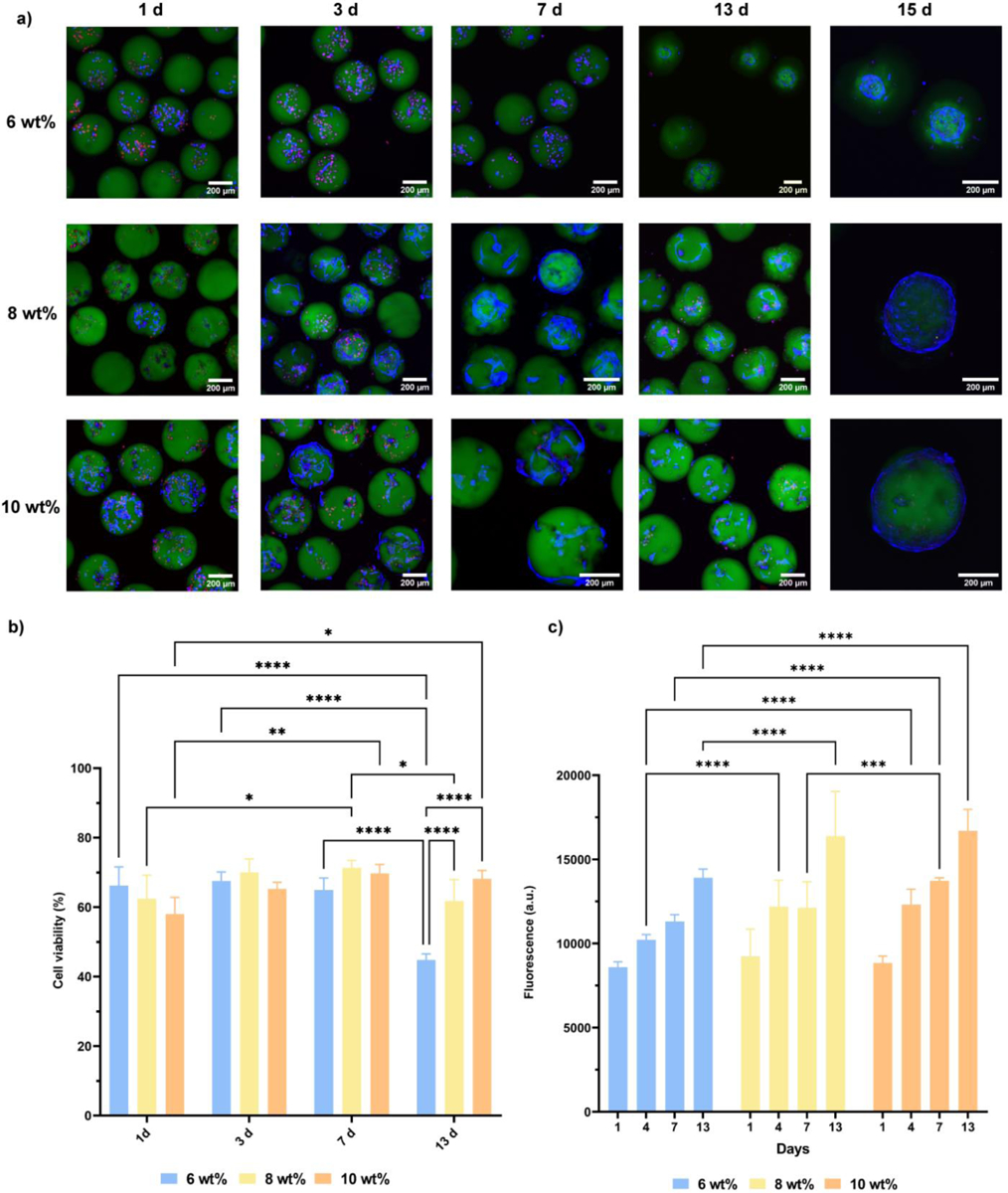
Figure 3:

Cell viability and metabolic activity of encapsulated hMSCs in 6, 8 and 10 wt% PEG-4aNB microgels over time. a) Confocal images of Live/Dead staining after 1, 3, 7, 13 and 15 days of culture. Live cells are shown in blue (LIVE 405 dye), dead cells in red (TOTO-3 dye) and microgels in green (PEG-FTIC). b) Cell viability analysis for encapsulated hMSCs in 6, 8 and 10 wt% PEG-4aNB microgels after 1, 3, 7 and 13 days of culture. c) Metabolic activity analyzed by alamarBlue assay of encapsulated hMSCs in PEG-4aNB microgels after 1, 3, 7 and 13 days of culture. n = 4 biologically independent samples, mean ± SD; *p < 0.01, **p < 0.001, ***p = 0.0002, ****p < 0.0001. Two-way ANOVA analysis was used to test for differences among groups.
