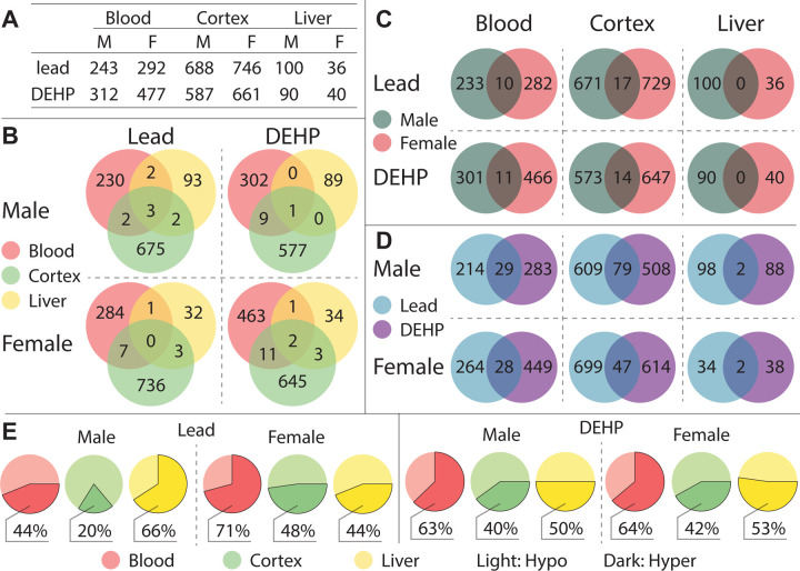
Figure 2: Summary of detected Differentially Methylated Regions.
Differentially Methylated Regions (DMRs) were categorized by tissue (blood, cortex, and liver), sex (F: female, M: male), and exposure group (Pb, DEHP, and control) (2A), and DMRs found in more than one tissue type were further categorized by sex and exposure (2B). DMRs shared by both sexes (2C) and by exposure group (2D) were quantified and broken down by tissue type. Proportions of DMR directional changes were generally summarized for each tissue-sex-exposure combination, designated by DNA hyper (more methylated) or hypo (less methylated), in comparison to controls (2E).

