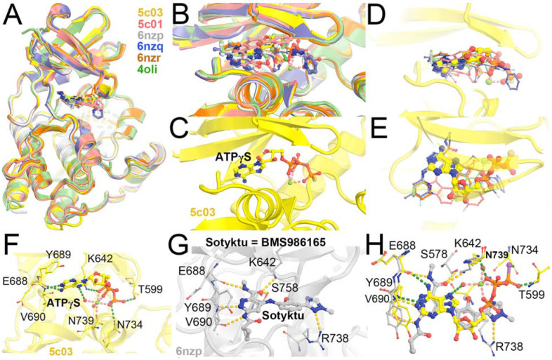
Figure 4.
Superposition of selected TYK2 JH2 complexes (PDB IDs 5c03, 5c01, 6nzp, 6nzq, 6znr, and 4oli). (A) Overall structure. (B) Closeup view of the binding pocket. (C) ATPγS-bound 5c03 complex. (D, E) Two orthogonal views of 5c03 structure with ATPγS and DEU (its three-letter residue code 2TT as used in the PDB) from 6nzp structure. (F) Detailed hydrogen bond interactions of ATPγS with TYK2 JH2 in 5c03 structure. (G) Detailed HB interactions of DEU/Sotyktu with TYK2 JH2 in 6nzp structure. (H) A combined view of ATPγS and DEU.

