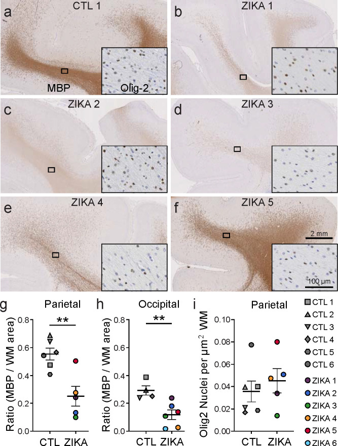
Fig. 2. Immunohistochemical analysis demonstrates marked reduction in myelin basic protein (MBP) in ZikV-exposed fetal NHP brains.
a-f, MBP (primary image) and Olig2 (inset) immunohistochemical staining of a) control and b-f) ZikV animals. The representative images are taken from dorsal parietal cortex; the black rectangle identifies the approximate location in the DWM tracts represented in the inset. g-h) Quantification of MBP staining in the DWM from g) parietal and h) occipital cortex, measured as the ratio of area occupied by chromogen divided by the total area of the DWM. i) Quantification of the density of Olig2+ nuclei within the DWM. Points in the plots represent individual animals (one slice per animal was quantified), with bars indicating mean ± SEM. **p<0.01 by unpaired t-test with Welch’s correction. Average gestational age (± SD) of ZikV-exposed vs CTL animals in IHC analysis=152(±2) vs 157(±9) days; p=0.23 by t-test.

