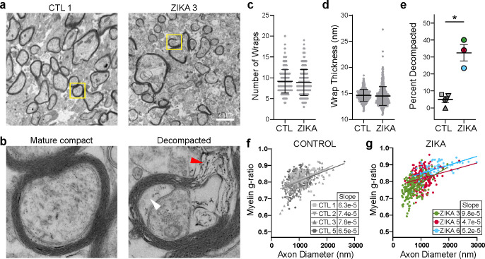
Fig. 4. ZikV exposure is associated with myelin sheath decompaction in fetal NHP brain.
EM analysis of brain sections sampled from DWM in parietal cortex. a-b) Representative EM images of myelinated axons from a control (left) and ZikV-exposed animal (right). The area marked by the yellow rectangle is expanded in panel b), demonstrating mature compact myelin (left) and decompacted myelin (right) with electron-dense material in the interlamellar space (red arrowhead) and swelling of the inter lamella (white arrowhead). c-d) Quantification of specific myelin features including the number of c) myelin wraps and d) average wrap thickness for CTL and ZikV animals, measured in areas of compact myelin. Individual points represent data for a single axon; error bars represent mean ± SEM across all points within a treatment condition. e) Percent of axons demonstrating a decompaction phenotype as defined by delamination of all layers of the myelin sheath with outward bowing, affecting at least 25% of the circumference of the axon. Individual points represent, for a single animal, the percent of myelinated axons with decompaction; error bars represent mean ± SEM across all points within a condition. *p<0.05. f-g) Plot of myelin g-ratio (outer diameter of axon divided by outer diameter of myelin sheath, measured in areas of compact myelin) versus axon diameter. Individual points represent a single myelinated axon. Linear regression was calculated for all points representing a single animal; slope is indicated in the table.

