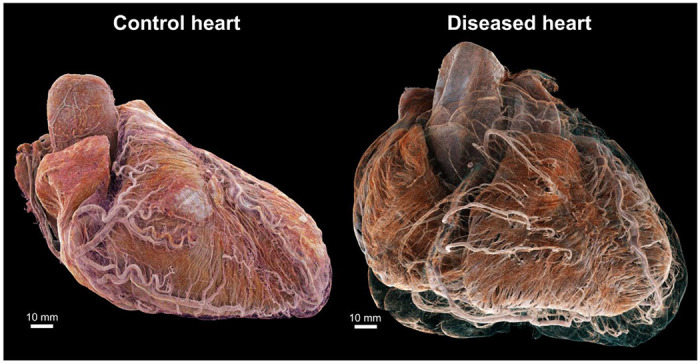
Fig. 1:

3D cinematic renderings of the normal (left) and abnormal (right) hearts in attitudinal position, i.e., as they would sit in the chest. The epicardial fat has been removed digitally to show the different course of the coronary vasculature between the two hearts. In the control, the coronary vasculature sits relatively close to the epicardial surface while in the disease heart, it is distant from the epicardium due to a thick layer of epicardial fat, while still being ‘anchored’ to the myocardium by smaller penetrating arteries. This gives rise to an unusual spiral configuration of the coronary arteries (see also Fig. 6g)
