Figure 4:

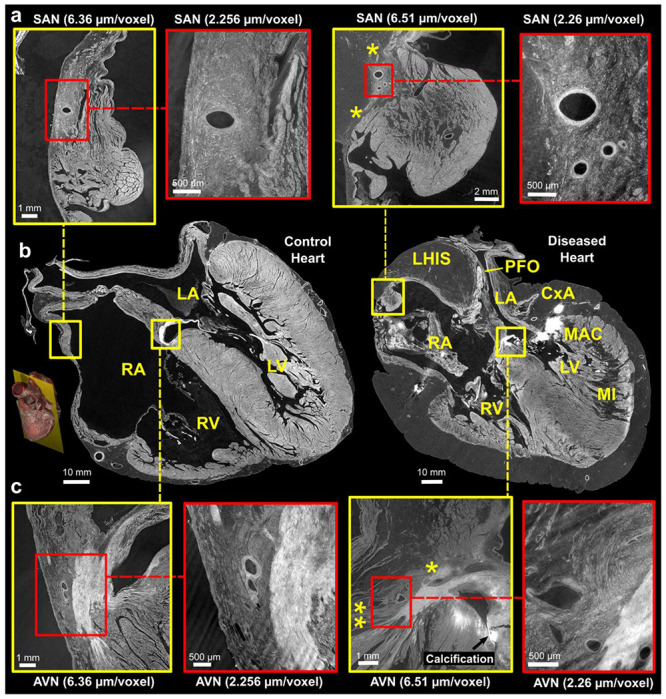
Comparison of control and pathological hearts - (b) Four-chamber slices of at an imaging resolution of 20 ^m/voxel, with locations of the sinus node (SN) and atrioventricular nodes (AVN) highlighted. A 3D rendering provides a visualization of the imaging plane in which these views were created. Zoom images of the SAN and A VN, taken from adjacent slices, are shown in (a) and (c), respectively. The yellow squares indicate images captured at a resolution of 6.5 μm/voxel, while the red squares indicate images captured at a higher resolution of 2.2 μm/voxel. The yellow stars in the diseased heart SAN indicate attenuated connections with working atrial myocardium running from the side of the SAN through epicardial fat and in AVN indicate connections of AVN with RA vestibule (**potential slow pathway) and attenuated connection with atrial septum (* potential fast pathway). LA - Left Atrium, RA - Right Atrium, LV - Left Ventricle, RV - Right Ventricle, MI - Myocardial Infarction, MAC - Mitral Annular Calcification, LHIS - Lipomatous Hypertrophy of the Inter-Atrial Septum, PFO - Patent Foramen Ovale. Note: The left atrium appears collapsed in both hearts.
