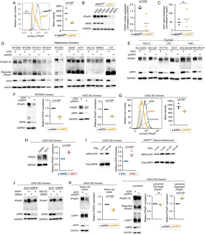
Figure 4: ARF6-dependent IFNγR1 surface expression in murine and human melanoma.
(A) Flow cytometric detection of IFNγR1 cell surface expression in early-passage murine tumor cell lines, n=3 independent cell lines. (B) Western blot for IFNγR1 in early-passage murine tumor cell lines, n=3 independent cell lines. (C) Quantitative RT-PCR for IFNγR1 in one early-passage murine tumor cell line, n=3 replicates. (D-F) Western blot for full length (FL) and degraded IFNγR1, ARF6 and GAPDH in human melanoma patient derived melanoma cell lines (MTG) and commercially available melanoma lines (D), in human non-small cell lung cancer (NSCLC), colorectal cancer (CRC), triple negative breast cancer (TNBC) cell lines (E) Quantification of IFNγR1 in MTG003 (n=3) and UACC.62 (n=3) cells (F) without or with ARF6 knockdown. (G) Flow cytometric detection of surface expression of IFNγR1 in UACC.62 cells without or with ARF6 knockdown, n=3. (H) Western blot for IFNγR1 in UACC.62 cells without or with 2μM QS11 treatment for 24hours, n=3. (I) Western blot for total ARF6 and ARF6 GTP in UACC.62 cells without or with 500U/mL IFNγ treatment, n=3. (J) Western blot for indicated proteins in UACC.62 cells without or with ARF6 knockdown and without or with 50nM Bafilomycin A1 or 10μM MG132 treatment for 6h. (K) Western blot analyses of UACC.62 cells without or with ARF6 knockdown as indicated, n=3. (A, B, C, G) t-test. (F, H, I, K) Ratio paired t-test. See also Figure S4.

