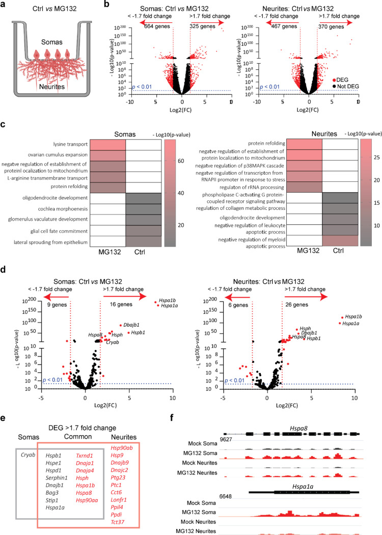
Fig. 1. Specific mRNAs are preferentially enriched in the soma or projections of hippocampal neurons after proteostatic stress.
(a) Schematic of primary mouse hippocampal neurons cultured in Transwell membranes to physically separate the soma and neurites for RNA extraction. Neurons were exposed to MG132 (10 μM for 7 h) or DMSO (Ctrl). (b) Volcano plot of differentially expressed genes (DEGs) in the soma or neurites (n = 3). Genes up- or down-regulated by > 1.7 fold after MG132 treatment and P-values < 0.01 are indicated in red. (c) Gene ontology enrichment analysis. Gene ontology categories of the top five biological processes enriched in DEGs in the somas and neurites after MG132 exposure. The color of the bands denotes the extent of upregulation. (d) Volcano plot of known chaperone-related genes. Genes up- or down-regulated by > 1.7 fold after MG132 treatment and P-values < 0.01 are indicated in red. (e) Venn diagram listing the differentially enriched molecular chaperone-related genes in the somas (gray square) and neurites (red square). (f) RNA-seq distributions of the Hspa8 and Hspa1a1 loci in the soma and neurites of control and MG132-exposed neurons.

