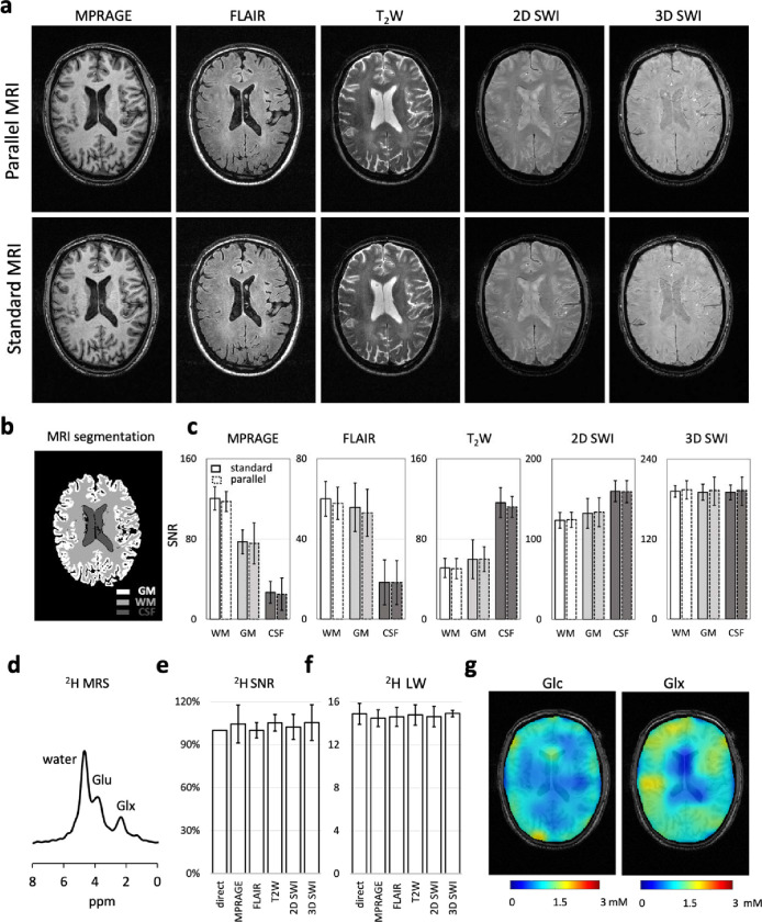
Figure 2. Parallel MRI-DMI on a healthy brain.
(a) Parallel and standard MP-RAGE, FLAIR, T2W, 2D SWI and 3D SWI MRIs acquired of a healthy brain. (b) The brain was segmented into white matter (WM), gray matter (GM) and cerebrospinal fluid (CFS) based on the MP-RAGE image. (c) The parallel versus standard MRI SNR comparisons in WM, GM and CSF are shown on the displayed brain slice. The error bars represent the standard deviation of image pixels(/voxels) in each segmented brain area. (d) The global 2H MR spectrum, acquired in parallel to the interleaved MRIs in (a), is shown. (e-f) The SNR and LW (of water peak) in the global 2H MR spectra are compared between the parallel and standard acquisitions (N=3). (g-h) DMI concentration maps of 2H-labeled Glc and Glx normalized to water, based on spectral fitting, assuming the concentration of deuterium in water remains constant across voxels (10 mM). One-way or two-way ANOVA were used for statistical analysis. No significant difference (P<0.05) was found between standard and parallel measurements in (c), (e) and (f).

