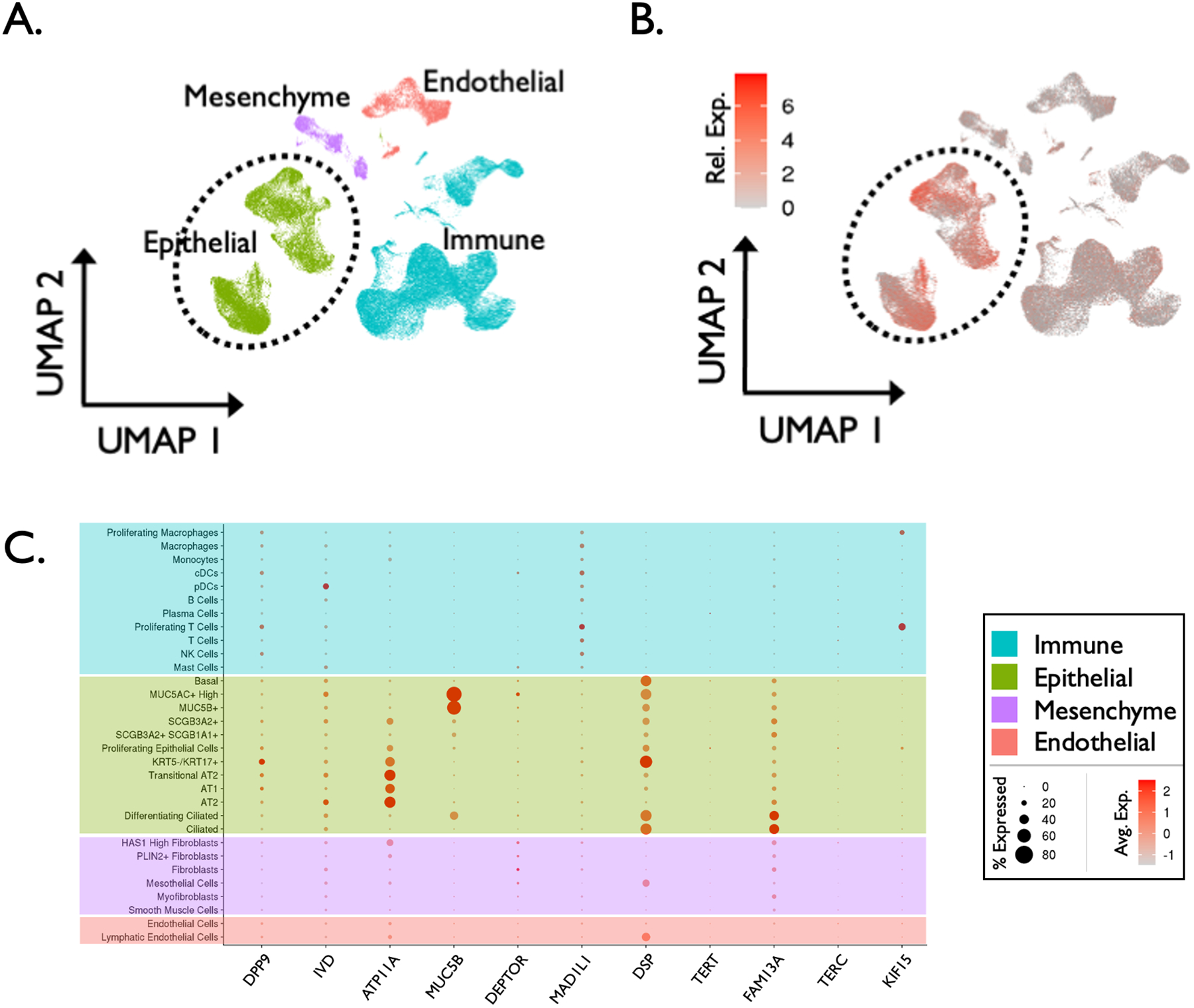
Figure 3.

Method validation: cell localization of idiopathic pulmonary fibrosis (IPF) candidate risk genes. (A) Single cell RNA sequencing (scRNA-seq) atlas comprised of 114,396 cells from 12 explanted IPF lung samples, clustered into 4 tissue types and visualized via UMAP projection. (B) Differential expression of 11 established IPF risk genes (RNA expression denoted by color gradient) among the 4 broad cell types demonstrating enriched expression of disease risk genes in epithelial cells in the IPF lung. (C) Additional dot plot depicting expression of 11 established IPF risk genes within 31 distinct cell types/states in the IPF lung (RNA expression denoted by color gradient, percentage of expressing cells within a cell type denoted by dot size).
