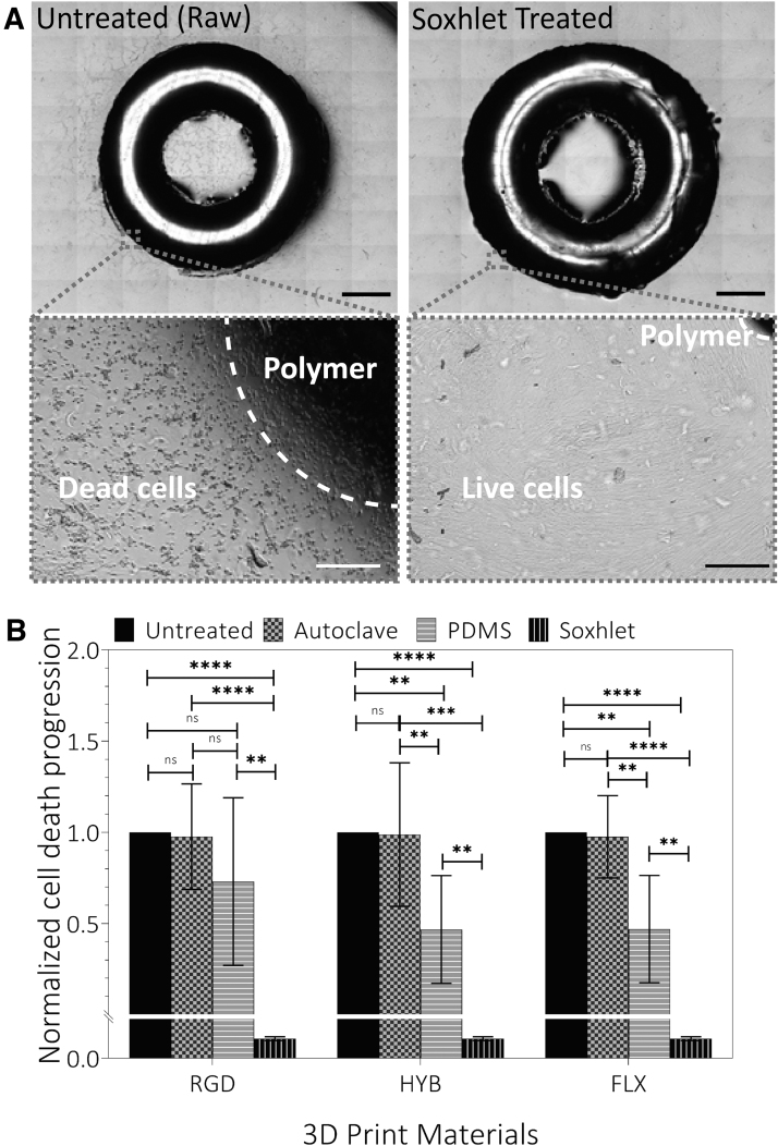
FIG. 3.
Cytotoxicity test of treated materials. (A) Representative stitched snapshots of the indirect contact cytotoxicity assay. Treated samples (Soxhlet extraction) showed drastically less cell death than the untreated (raw) sample. Darker zones indicate the areas of cell death caused by cytotoxicity of the material, while lighter zones show live cells. Both snapshots show RGD material on L929 cells after 36 h. The blowup images (bottom) show typical apoptotic (shrink) and healthy cell morphologies that led to dark and bright tones, respectively, in the stitched images. Scale bars: Top: 2 mm; Bottom: 0.1 mm. (B) Progression of cell death after 36 h, measured as the distance from each treated and untreated ring, showing little detoxification with autoclaved samples, partial detoxification with PDMS-coated samples, and complete detoxification with Soxhlet extracted samples. The areas of cell death were normalized to the control. Note: Y-axis was segmented to show the Soxhlet extraction value as it was close to zero. Statistical analysis: Student t-test. **p < 0.01; ***p < 0.001; ****p < 0.0001. Sample size: Three samples were recorded in eight different locations each. NS, not significant; PDMS, polydimethylsiloxane.

