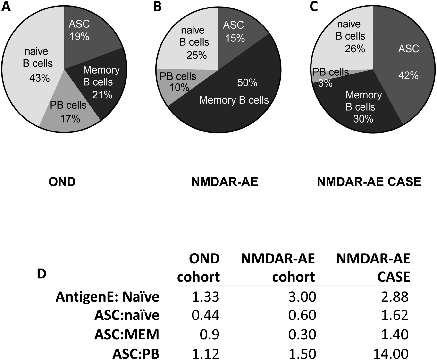
FIGURE 1. B cell profile in the CSF of pediatric Autoimmune Encephalitis.

Cerebrospinal fluid (CSF) was collected from pediatric patients with autoimmune encephalomyelitis (AE). Shown are B cell subset frequencies by flow cytometry for (A) pediatric patients who tested negative for antibodies against the NMDAR (OND), (B) pediatric patients who tested positive for antibodies against the NMDA receptor and met diagnostic criteria for NMDAR-AE (NMDAR-AE) and (C) one pediatric NMDAR-AE case refractory to treatment (NMDAR-AE CASE). (D) Ratios of B cell subsets in each group. In D, abbreviations are as follows: AntigenE, Antigen Experienced; ASC, antibody secreting cell; MEM, memory; PB, plasmablast.
