
FIGURE 5: Comparison of rhAbs to germline configuration.
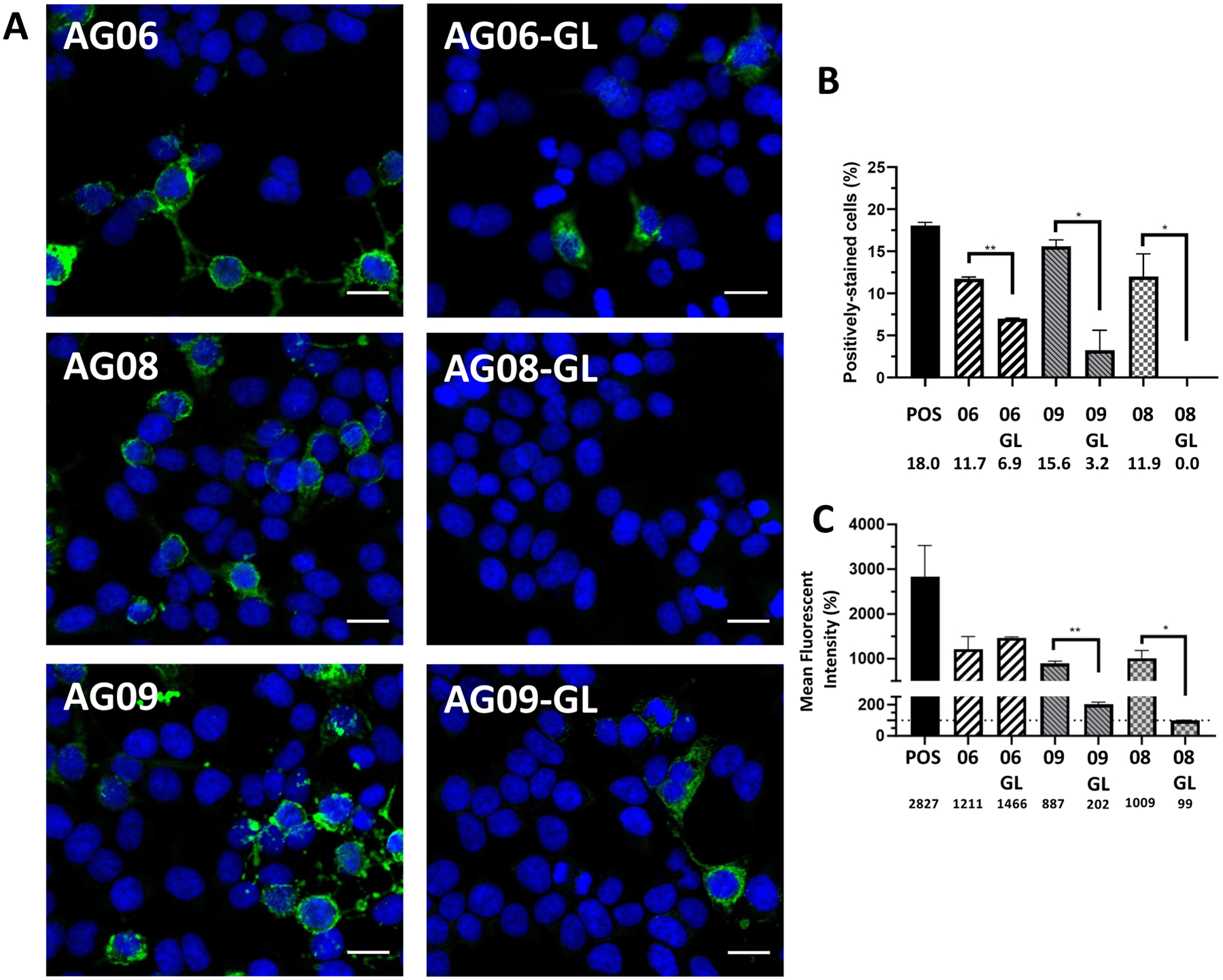
(A) Antibodies expressed by individual B cells from the CSF of a pediatric NMDAR-AE patient refractory to treatment were reverted to their germline configuration and tested for binding to the NR1 subunit of the NMDAR using a commercially available kit as described in the methods. Blue: DAPI staining. Green: NMDAR. Scale bar: 20 μm. (B) Frequency of positively-stained cells. (C) Mean fluorescence intensity of cells. The horizontal dotted line represents the MFI of control cells as the threshold of detection.