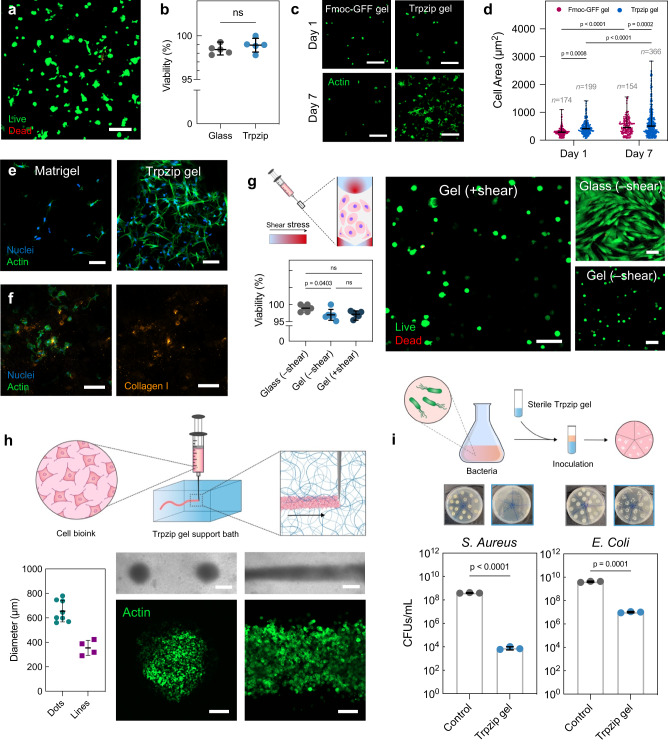
Fig. 4. Trpzip hydrogels support cell growth, syringe extrusion, biofabrication and show antimicrobial properties.
a Confocal microscopy image of human fibroblast cells stained with Calcein AM and ethidium homodimer in Trpzip hydrogels (1% w/v, DMEM, pH 7) after 5 days in culture. The scale is 100 μm. b Quantified percentage of cell viability of fibroblasts cultured in Trpzip hydrogels compared to culture on glass (n = 5 gels). Data are presented as mean ± s.d. P-values calculated using two-tailed unpaired t-test. c Confocal microscopy image of fibroblasts cultured in Fmoc-GFF hydrogels (1% w/v) and Trpzip hydrogels (1% w/v) after seven days, without the use of RGD binding ligands. The scale bar is 50 μm. d Quantification of cell area (left) and cell aspect ratio (right) for fibroblasts cultured in Fmoc-GFF hydrogels (n = 3) and Trpzip hydrogels (n = 3) for seven days. Error bars show mean ± s.d. P-values calculated using two-way ANOVA. e Confocal microscopy image of HFFs cultured in Matrigel and Trpzip gels (no RGD) for 30 days. The scale bar is 100 μm. Data is representative of one independent experiment. f Deposition of endogenous collagen I by fibroblast cells after 14 days in culture in Trpzip hydrogels without RGD ligands. The scale bar is 100 μm. Data is representative of one independent experiment. g Viability of cells encapsulated in Trpzip hydrogels and then exposed to shear (n = 3 gels) compared to cells experiencing no shear seeded on glass (2D; n = 3 wells) or within Trpzip gels (3D; n = 3). Error bars show mean ± s.d. P-values calculated using one-way ANOVA. The scale is 100 μm. h Bioprinted constructs of cells in a Trpzip hydrogel support bath, either as droplets (left panel) or lines (right panel). The scale is 500 μm in optical images and 200 μm in immunofluorescence images. The measured average diameter of printed cellular droplets (n = 8) or lines (n = 4). Error bars show mean ± s.d. i Antibacterial activity of Trpzip hydrogels against both a gram positive (S. aureus) and a gram negative (E. coli) species, assessed using a bacterial growth inhibition assay. Photographs show agar plates used to count colony forming units (CFUs) per mL of each bacterial species after incubation with Trpzip gels for 24 h at 37 °C (n = 3 gels). Data are presented as mean ± s.d. P-values calculated using two-tailed unpaired t-test.

