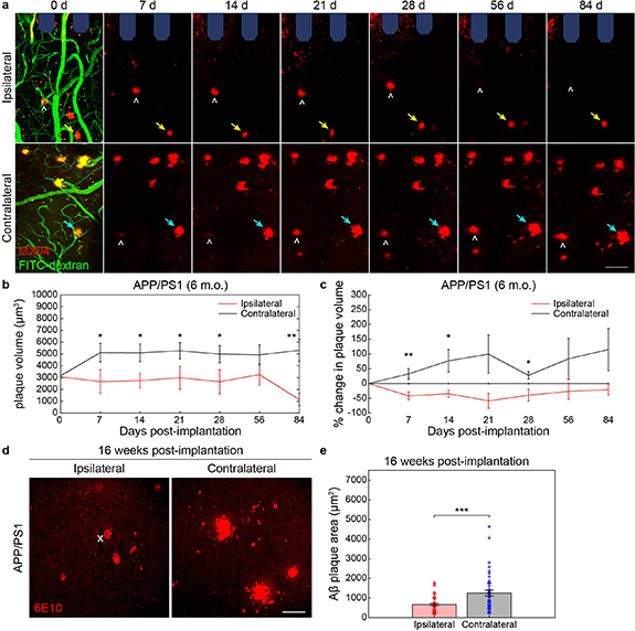
Figure 3.

Chronic microelectrode implantation reduces the growth of local amyloid plaques in adult APP/PS1 mice. (a) Representative two-photon images of methoxy-X04 labeled plaques (MX04, red) and blood vessels (FITC-dextran, green) in ipsilateral hemisphere around multi-shank microelectrode array (shaded blue) over 12 weeks post-implantation in adult (6 m.o.) APP/PS1 mice compared to contralateral (uninjured) hemisphere. Ipsilateral hemisphere demonstrates plaques which do not visually change in size with chronic implantation (yellow arrow) whereas plaques on the contralateral hemisphere appear to increase in size over time (cyan arrow). NOTE: some plaques move into and out of frame over time (white hat) due to tissue drift between subsequent chronic imaging sessions. Scale bar = 50 μm. (b) Change in plaque volume over a 12 week implantation period between ipsilateral and contralateral hemispheres in adult APP/PS1 mice (25 Aβ plaques on ipsilateral hemisphere and 27 plaques on contralateral hemisphere tracked longitudinally over seven time points across n= 3 mice). (c) Percent change in plaque volume with respect to plaque size on day 0 of electrode insertion over a 12 week implantation period between ipsilateral and contralateral hemispheres in adult APP/PS1 mice. (d) Representative immunohistology stain for 6E10, an Aβ marker, following 16 weeks post-implantation in adult (6 m.o.) APP/PS1 mice demonstrating visually reduced Aβ plaque sizes in ipsilateral hemisphere around the site of probe insertion (denoted by white ‘x’) compared to contralateral side. Scale bar = 100 μm. (e) Average Aβ plaque area measured by 6E10 stain between ipsilateral and contralateral hemispheres (n= 42 Aβ plaques on ipsilateral hemisphere over 13 histological tissue sections and n = 45 Aβ plaques on contralateral hemisphere over 17 histological sections across 6 mice total). * p < 0.05, ** p < 0.01, *** p < 0.001. All data is reported as mean ± SEM.
