Figure 6.

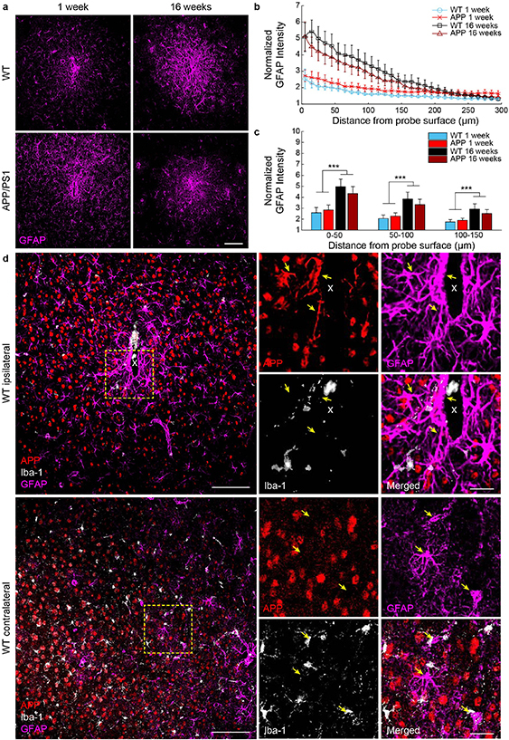
Reactive astrocytes express amyloid precursor protein around chronically implanted microelectrode arrays at 1 and 16 weeks post-implantation in WT and APP/PS1 mice. (a) Representative images of GFAP+ reactive astrocyte staining (magenta) around implanted microelectrodes. Scale bar = 100 μm. (b) Normalized GFAP fluorescence intensity with respect to distance from the implanted microelectrode. (c) Average GFAP fluorescence intensity within 50 μm bins up to 150 μm around chronically implanted microelectrodes (n= 6 mice per group at 1 week, n= 7 mice per group at 16 weeks). (d) Immunohistological example demonstrating expression of amyloid precursor protein (APP, red) in GFAP+ astrocytes (magenta) but not Iba-1 + microglia (white) on the ipsilateral, but not contralateral hemisphere, near the site of a chronically implanted microelectrode in a WT mouse. Scale bar = 100 μm, 25 μm (inset). *** p < 0.001. All data is reported as mean ± SEM.
