Figure 5.
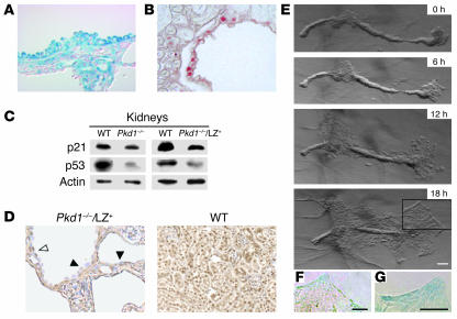
Proliferation of Pkd1–/– cyst epithelial cells. (A) A kidney from a P17 Pkd1–/–/LZ+ mouse was stained with β-gal and counterstained with Nuclear Fast Red. LZ+ epithelial cells occasionally showed focal hyperplastic features such as micropolyps. Original magnification, ×400. (B) A kidney froma P12 Pkd1–/–/LZ+ mouse was stained with an anti-PCNA. Some cuboidal LZ+ cyst epithelial cells were accompanied by PCNA expression. Original magnification, ×400. (C) Expression of p21 and p53 in the kidneys of Pkd1–/– mice at E16.5 and Pkd1–/–/LZ+ mice 1 month of age. The amount of p21 and p53 in kidneys was examined using Western blot. Actin was used as a loading control for protein. Data presented are 1 representative of 4 independent experiments. (D) Kidneys of P12 Pkd1–/–/LZ+ and P12 wild-type mice were stained with anti-p53. Expression of p53 was detected in the flat epithelial cells (white arrowhead) but was significantly decreased in the cuboidal cyst epithelial cells (black arrowheads) of the Pkd1–/–/LZ+ mouse. Original magnification, ×400. (E–G) The proliferation of cyst epithelial cells in vitro. A single nephron isolated by microdissection from the kidney of a Pkd1–/–/LZ+ mouse at E17.5 was cultured in collagen gel for 18 hours. (F and G) Higher magnifications of the boxed area above. Both LZ+ and Pkd1–/– (LZ–) cells proliferated. Scale bars: 100 μm.

