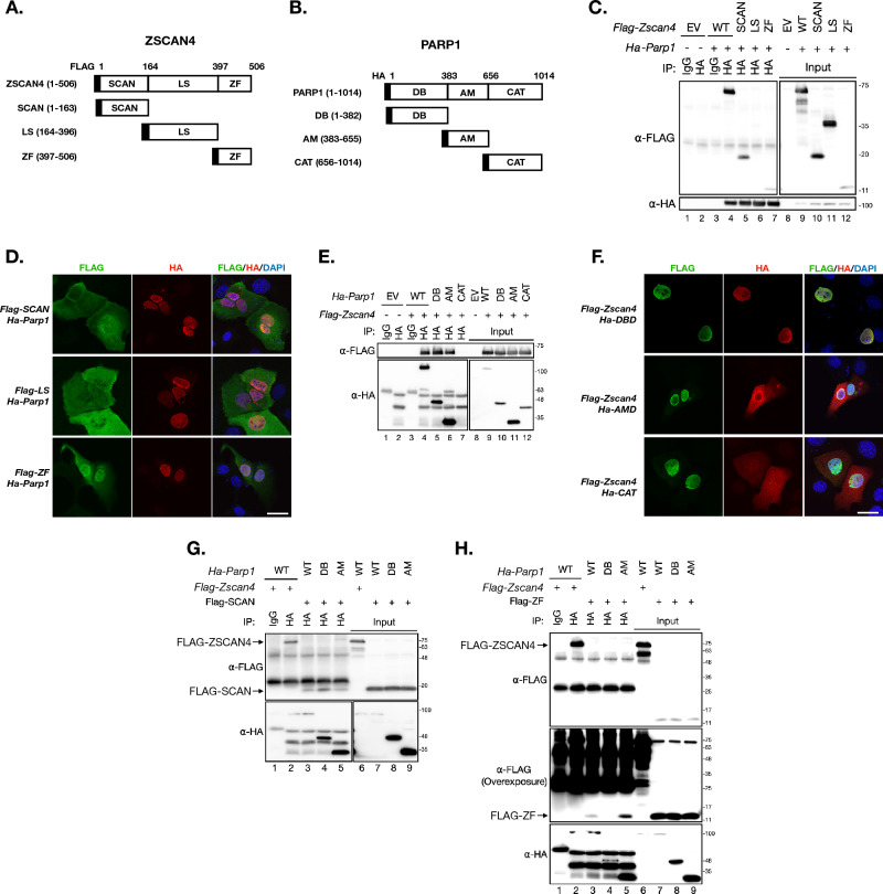
Fig. 4.
ZSCAN4 and PARP1 interacts with each other. A The illustration of ZSCAN4 protein domains. The numbers on the top indicate the residue positions. SCAN: the SCAN domain (amino acid residue 1–163 of ZSCAN4); LS: the linker sequence (amino acid residue 164–396 of ZSCAN4); ZF: the zinc finger domain (amino acid residue 397–506 of ZSCAN4). B The illustration of PARP1 protein domains. DB: the DNA binding domain (amino acid residue 1–382 of PARP1); AM: the auto-modification domain (amino acid residue 383–655 of PARP1); CAT: the catalytic domain (amino acid residue 656–1014 of PARP1). C Co-IP of individual FLAG-ZSCAN4 domains (SCAN, LS or ZF) and full-length HA-PARP1. D IF images of FLAG (indicative of ZSCAN4 domains) and HA (indicative of full length PARP1) in mouse BNL CL.2 transiently expressing full length HA-PARP1 and a FLAG tagged ZSCAN4 domain. Scale bar: 25 µm. E Co-IP of individual HA-PARP1 domains (DB, AM or CAT) and full-length FLAG-ZSCAN4. F IF images of FLAG (indicative of full length ZSCAN4) and HA (indicative of PARP1 domains) in mouse BNL CL.2 transiently expressing full length FLAG-ZSCAN4 and a HA tagged PARP1 domain. Scale bar: 25 µm. G Co-IP of the HA-DB and HA-AM (of PARP1) with FLAG-SCAN (of ZSCAN4). The top arrow indicates the full-length FLAG-ZSCAN4 bands. The lower arrow indicates the FLAG-SCAN domain (of ZSCAN4) bands. WT: wildtype. H Co-IP results of the HA-DB and HA-AM (of PARP1) with FLAG-ZF (of ZSCAN4). The top arrow indicates the full-length FLAG-ZSCAN4 bands. The lower arrow indicates the FLAG-ZF domain (of ZSCAN4) bands. The middle panel is an overexposure of the top panel to reveal the FLAG-ZF bands. WT: wildtype

