Figure 1.
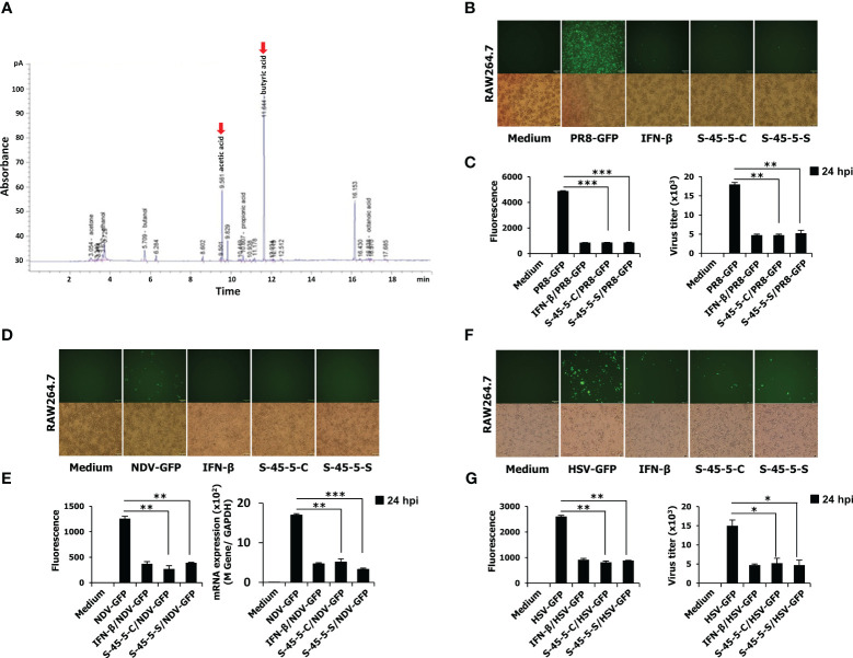
The organic acid production profile of C. butyricum S-45-5 and its antiviral activity in RAW264.7 cells. (A) HPLC analysis was carried out by using C. butyricum S-45-5 culture supernatant as the sample and ethanol, acetic acid, butanol, and butyric acid as standards. Employing helium as the carrier gas at a flowrate of 1 ml/min, the oven temperature was stepwise elevated from 50°C to 170°C at a rate of 10°C/min. The injector and detector temperatures were set to 250°C. RAW264.7 cells were added into 12-well cell culture plates, each well containing 2.5 × 10⁵ cells/well. Twelve hours later, cells were treated with PBS (medium, virus only), or 100 U/ml rmIFN-β, or C. butyricum S-45-5-Cell (1 × 10⁶ CFU/ml) or S-45-5-Sup (1 × 10⁶ CFU/ml). The medium was replaced with 1% fetal bovine serum (FBS) in Dulbecco’s modified Eagle’s medium (DMEM) 12 h later, and except for the medium, all other cells were infected with the green fluorescent protein (GFP) fused (B, C) H1N1 influenza virus (PR8-GFP, 1 MOI), (D, E) Newcastle disease virus (NDV-GFP, 2 MOI), or (F, G) Herpes simplex virus (HSV-GFP, 0.5 MOI). After 2 h, 10% FBS containing DMEM was added to the medium, and images were obtained following 24 h (200× magnification). GFP absorbance levels were measured by the GloMax multi-detection luminometer (Promega). A plaque assay was used to measure the virus titration in the supernatant and cells, and plaque-forming units were expressed (PFU). Clostridium butyricum S-45-5-Cell (S-45-5-C) and Clostridium butyricum S-45-5-Sup (S-45-5-S). GFP absorbance and virus titer are expressed as mean ± standard deviations (SD). Error bars indicate the range of values obtained from counting duplicates in three independent technical experiments (*p < 0.05, **p < 0.01, and ***p < 0.001 regarded as significant differences).

