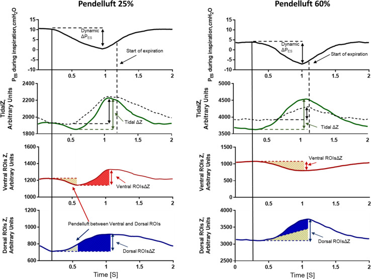
Figure 1.
Representative figure of two patients (left and right panels) during the trial, showing pendelluft between the ventral and dorsal regions of the lung. Pendelluft percentage was calculated as described in the Methods and the online supplement. In both patients, during muscular inspiration (first panel), air moves from the ventral regions of interest (ROIs) (third panel) toward the dorsal ROIs (fourth panel) and vice versa during muscular expiration. This pendelluft effect is represented as the yellow area that moves from the ventral ROIs (third panel) to the dorsal ROIs (fourth panel). The second panel shows tidal impedance variation (TidalΔZ, a surrogate of Vt) computed as: 1) the difference between the end-expiratory and end-inspiratory impedance during the breathing cycle (dashed line); and 2) the gas inflating the lungs during the breathing cycle, calculated on a pixel-by-pixel basis (solid green line). TidalΔZ, computed as the difference between the end-expiratory and end-inspiratory impedance (dashed line), does not consider the intratidal shift between the ventral and dorsal ROIs. This yields underestimation of the volume of lung distension, especially in the dorsal regions. TidalΔZ, calculated on a pixel-by-pixel basis (solid green line), represents a more accurate estimate of the amount of lung distension. A detailed description of the TidalΔZ calculation is provided in the online supplement. PES = esophageal pressure; ROIsΔZ = tidal impedance variation within ROIs; ΔPES = inspiratory effort.

