FIG 2.
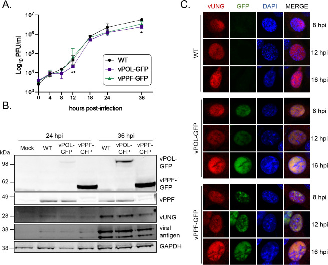
Characterization and immunofluorescence imaging of recombinant MHV68 expressing GFP-tagged vPOL and vPPF. (A) Single-step growth curve to compare replication of wild-type MHV68 (WT) or recombinant MHV68 expressing GFP fusions to vPOL (viral DNA polymerase encoded by ORF9) or vPPF (viral DNA polymerase processivity factor encoded by ORF59) in NIH 3T12 fibroblasts, MOI 3.0. Symbols represent three biological replicates ±SD. Statistical significance was evaluated by two-way ANOVA with Tukey’s multiple comparisons test, and differences were noted between ORF9-GFP and WT at 12 and 36 hpi and between ORF9-GFP and ORF59-GFP at 12 hpi. *, P < 0.05; **, P < 0.01 (B) Timecourse of viral protein expression by immunoblot under conditions described in A. (C) Immunofluorescence timecourse of viral protein co-localization in NIH 3T3 cells infected with the indicated viruses, MOI 3.0. vUNG was detected with vUNG pAb, followed by secondary AF568; vPOL and vPPF fusion proteins were detected via GFP expression without antibodies; DNA was stained with DAPI.

