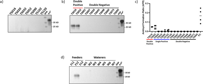
Fig 2.
Representative results of western blot, PMCA, and RT-QuIC screening on exhumed tongue samples. (a) Representative samples displaying negative PrPSc-associated signals in western blot after proteinase K treatment. (b) PMCA analyses in exhumed tongue tissues. Results in this panel correspond to a fourth PMCA round, and they depict eight samples displaying negative signals in both replicates (“Double −”) and the two samples that were positive for PMCA in both replicates (“Double +”). The PMCA procedure was performed as described in Ref.13 with no modifications besides the use of tongue tissue. Codes at the top of panels (a) and (b) represent codes from individual animals. (c) Representative RT-QuIC data including all PMCA-positive samples and eight representative samples that provided PMCA-negative results in both replicates. “PC” represents RT-QuIC data of a retropharyngeal lymph node from a pre-clinical white-tailed deer used as a positive control. (d) PMCA analysis of representative feeders (F11, F12, F13) and waterers (W1–W7) swabs. As in (b), these results also correspond to a fourth PMCA round. Numbers at the right of panels (a), (b), and (d) correspond to molecular weight (MW) markers. All samples in (a), (b), and (d) were treated with proteinase K with the exception of “PrPC” which corresponds to the brain extract of tg1536 mice (used to prepare PMCA substrate) that is utilized as an electrophoretic mobility and antibody specificity control. All blots were developed using the 8H4 antibody.

