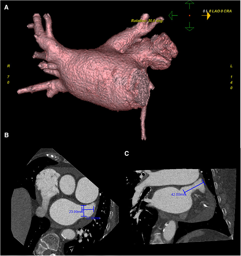
Figure 1.
Preoperative cardiac CT angiography reveals a complex left atrial appendage with a large branch, no clear filling defect in the left atrial appendage and left atrium, and no diverticulum and accessory atrial appendage was identified in the left atrium (A) the cross-sectional image of the LAA orifice (B) the long diameter was about 2.4 cm, the short was about 2.2 cm. Oblique view (C) the maximum depth of the left atrial appendage branch was about 4.2 cm. Pericardial effusion.

